Time To Read
24–36 minutes
Date Last Modified
Print your own flashcards
Pick the terms you want and choose a printout — fold-over cards, cut-apart double-sided cards, index cards, a two-column study sheet, or a fill-in-the-blank quiz. The terms come straight from the glossary below, so they always match the course.
Open the flashcard maker in a new page — easier on a phone, and better if you use a screen reader or keyboard only.
Muscle Origins, Insertions & Actions
Each muscle is listed with where it starts, where it ends and what it does, grouped by body region. These are drawn from the individual muscle pages on this site.
Abdominal muscles
- External Oblique – Origin: The rectus abdominis originates from the pubic symphysisCartilaginous joint connecting the two pubic bones at the midline. and the pubic crestRidge on superior surface of pubic body; attachment for abdominal muscles.. Insertion: The rectus abdominis extends upwards along the midline of the abdomen. It inserts into the xiphoid processSmall, cartilaginous tip at the inferior end of the sternum; often pointed. of the sternumFlat bone forming the center of the chest; anchor for ribs and clavicles. Appears as a vertical dagg and into the costal cartilages of the fifth, sixth, and seventh ribsCurved bones forming the rib cage; articulate with thoracic vertebrae and most with the sternum.. Actions: Posture and Movement; Trunk Flexion.
- Internal Oblique – Origin: The internal abdominal obliques originate from several points: the inguinal ligament, iliac crestSuperior ridge of the ilium; easily felt along the waistline., and thoracolumbar fascia. They also have attachments to the lower three or four ribs. Insertion: Fibers converge and attach to the linea alba. They also attach to the pubic crest, the xiphoid process of the sternum, and the cartilages of the lower three or four ribs. Actions: Posture and Movement; Lateral Flexion; Trunk Rotation and Flexion.
- Rectus Abdominis – Origin: The rectus abdominis originates from the pubic symphysis and the pubic crest. Insertion: The rectus abdominis extends upwards along the midline of the abdomen. It inserts into the xiphoid process of the sternum and into the costal cartilages of the fifth, sixth, and seventh ribs. Actions: Posture and Movement; Trunk Flexion.
- Transverse Abdominis – Origin: The transversus abdominis muscle originates from the lower six costal cartilages, the anteriorThe front of the body or toward the front when standing in the anatomical position. two-thirds of the iliac crest, and the lateralAway from the midline of the body. third of the inguinal ligament. Its origin is on the inner surface of the lower ribs, the lumbar vertebraeLargest and thickest vertebrae; short, blunt spinous processes for weight-bearing stability., and the pelvis. Insertion: The muscle inserts into the linea alba, the pubic crest, and the pecten pubisAnterior portion of the hip bone; meets its pair at the pubic symphysis.. Actions: Posture and Movement; Pelvic Stability; Spinal Support.
Anterior Thigh muscles
- Adductor longus – Origin: The adductor longusAdductor On the inner thigh; pulls the leg toward the midline muscle originates from the pubic bone. It specifically originates from the pubic crest. The pubic crest is a ridge on the superior borderTop edge of the scapula; shortest of the three borders. of the pubic bone. Insertion: The muscle inserts into the middle third of the linea asperaProminent ridge running down posterior shaft; attachment for thigh muscles.. This is a ridge on the posterior surface of the femurThigh bone; longest and strongest bone in the body; has a large round head and prominent trochanters (thigh bone). The insertion helps to stabilize the femur during movements of the hip joint. Actions: Hip Adduction Assistance in Hip Flexion and Extension Stabilization of the Hip Join.
- Gracilis – Origin: The gracilis muscle originates from the pubic symphysis. It is a cartilaginous joint at the midline of the pelvis. It also originates from the pubic crest, which is a bony ridge in the pelvic bone. Insertion: The gracilis muscle attaches to the medialToward the midline of the body surface of the tibiaShinbone; large, weight-bearing medial bone of the lower leg.. It specifically connects to the proximalCloser to the point of attachment or origin. part of the tibia, just below the knee joint. It inserts via a strong tendon that becomes part of the pes anserinus, a tendinous insertion involving three muscles. Actions: Hip Adduction Knee Flexion Assistance in Medial Rotation of the Knee.
- Illiacus – Origin: The iliacusFlexor Inside the hip bone; joins with psoas to flex the thigh. muscle originates from the iliac fossaLarge, smooth concave surface on the internal (medial) side of the ilium.. This is a concave, smooth surface on the inner aspect of the pelvic bone. This bone is called the iliumBroad, flaring upper portion of the hip bone; forms the “wings” of the pelvis.. Specifically, the iliacus arises from the upper two-thirds of the iliac fossa, which is part of the pelvis. Insertion: The muscle fibers of the iliacus converge and extend downward. They insert medially onto the femur, specifically the lesser trochanterSmaller medial projection below the head; muscle attachment site.. The lesser trochanter is a bony prominence on the proximal part of the femur. It is located on the medial side, just below the greater trochanterLarge lateral projection near the head; palpable on the side of the hip.. Actions: Hip Flexion Stabilization of the Hip Joint Assistance in Lumbar Spine Flexion.
- Psoas – Origin: The psoas majorFlexor Runs from lower spine to femur; lifts your thigh or bends your trunk. muscle has two primary origins. It originates from the bodies and intervertebral discs of the lumbar vertebrae, specifically T12 to L5. It also comes from the transverse processes of the lumbar vertebrae. Additionally, it arises from the lateral aspects of the intervertebral discs. It also originates from the adjacent vertebral bodies and the transverse processes of the lumbar vertebrae. Insertion: The psoas major muscle inserts on the lesser trochanter of the femur. The muscle fibers extend downward and laterally. They converge into a tendon that inserts onto the posterior surface of the lesser trochanter of the femur. This insertion point allows the psoas major to play a crucial role in hip flexion and stability. Actions: Hip Flexion Lumbar Spine Stabilization Lateral Flexion of the Spine.
- Rectus femoris – Origin: The anterior inferior iliac spineSmall bump below the ASIS; site for muscle attachment. (AIIS), which is a bony projection at the front of the hip bone (ilium). The acetabulumDeep, cup-like socket on lateral side where femur head articulates; shared by all three hip bone reg, which is the cup-like socket in the hip bone where the femur (thigh bone) inserts. Insertion: The rectus femorisExtensor / Flexor Front of thigh; straightens the knee and helps lift the leg at the hip. inserts via the quadriceps tendonConnector Above patella; connects thigh muscles to kneecap. into the patellaKneecap; small triangular bone embedded in quadriceps tendon; protects knee joint. (kneecap). From the patella, the quadriceps tendon continues as the patellar ligament. It attaches to the tibial tuberosityRough bump below kneecap on anterior surface for patellar ligament attachment. on the anterior surface of the tibia (shinbone). Actions: Knee Extension Hip Flexion Assistance in Trunk Flexion.
- Sartorius – Origin: The sartoriusFlexor / Rotator Runs diagonally from outer hip to inner knee; flexes, abducts, and rotates the thig muscle originates from the anterior superior iliac spineProminent front projection of the iliac crest; common landmark for muscle attachment. (ASIS) of the pelvis. Insertion: It inserts into the medial surface of the tibia. More specifically, it attaches at the area just below the medial condyleInner rounded bump at distal end; articulates with tibia and meniscus. of the tibia. This region is known as the pes anserinus. Actions: Hip Flexion Hip Abduction External Rotation.
- Tensor fascia latae – Origin: The tensor fascia lata starts from the anterior superiorAbove or toward the upper part of the body. iliac spineProminent ridge on the posterior scapula dividing it into supraspinous and infraspinous fossae. (ASIS). It also originates from the outer lip of the iliac crest, just below the ASIS. The ASIS is a prominent bony projection on the anterior part of the pelvis. It serves as a key landmark for various muscles and ligaments in the region. Insertion: From its origin, the TFL travels downward and slightly backward. It merges with the iliotibial band (IT band). The IT band is a thick band of connective tissue. It extends down the lateral side of the thigh. The TFL inserts indirectly via the IT band into the lateral condyleOuter rounded bump at distal end; articulates with tibia and meniscus. of the tibia, specifically at Gerdy’s tubercle. This insertion allows the TFL to influence movements at both the hip and knee joints. This makes it a versatile and functional muscle. Actions: Hip Flexion Hip Abduction Medial Rotation of the Hip.
- Vastus lateralis – Origin: The vastus lateralisExtensor Outer thigh muscle; extends knee and supports patella movement. muscle originates from the lateral lip of the linea aspera. This is a ridge on the posterior surface of the femur. It also has origins from the greater trochanter and the intertrochanteric line of the femur. Insertion: The vastus lateralis inserts into the quadriceps tendon, which then merges with the patella. From the patella, the quadriceps tendon continues as the patellar ligament. It attaches to the tibial tuberosity on the anterior surface of the tibia. Actions: Knee Extension Assistance in Hip Flexion Stabilization of the Patella.
- Vastus medialis – Origin: The vastus medialisExtensor Inside part of thigh; extends knee and stabilizes the kneecap. originates from the lower part of the femur. It specifically comes from the intertrochanteric line and the upper part of the linea aspera. The muscle fibers extend downwards along the femoral shaft. Insertion: The vastus medialis inserts into the patella (kneecap) via the quadriceps tendon. From the patella, the quadriceps tendon continues as the patellar ligament. This ligament inserts into the tibial tuberosity on the anterior surface of the tibia. Actions: Knee Extension Stabilization of the Patella.
Back muscles
- Latissimus dorsi – Origin: The latissimus dorsiExtensor / Adductor Covers lower back; pulls arms down and back, ‘swimmer’s muscle’. muscle has a wide origin. It primarily originates from the thoracic and lumbar vertebrae. It also comes from the lower ribs and iliac crest (part of the pelvic bone). Insertion: The muscle fibers of the latissimus dorsi converge. They insert into the floor of the intertubercular groove of the humerusLong bone of the upper arm; articulates with scapula at shoulder and radius/ulna at elbow.. This provides a solid attachment to the upper arm bone. Latissimus dorsi Actions Shoulder Extension. Actions: Shoulder extension; adduction of the arm; medial rotation of the arm.
- Rhomboid major – Origin: The rhomboid majorRetractor Below rhomboid minor; also draws shoulder blades inward. muscle originates from the spinous processes of the T2 to T5 thoracic vertebraeHeart-shaped body, long downward spinous process, and facets for rib attachment.. Insertion: Both rhomboid major and minor muscles insert onto the medial borderLong edge parallel to the vertebral column (closer to spine). of the scapulaTriangular “shoulder blade” on posterior thorax; connects clavicle and humerus.. The rhomboid major inserts from the root of the spine of the scapula to the inferior angleBottom tip of scapula where medial and lateral borders meet; visible when arm is raised.. The rhomboid minorRetractor Upper back; pulls shoulder blades toward the spine. inserts slightly above it. Rhomboid major Actions Shoulder Elevation. Actions: Shoulder elevation; shoulder girdle stability; scapular retraction.
- Rhomboid minor – Origin: The rhomboid minor muscle originates from the spinous processPosterior projection from vertebral arch; palpable bumps down the back. of the C7 and T1 vertebrae. Insertion: Both rhomboid major and minor muscles insert onto the medial border of the scapula. The rhomboid major inserts from the root of the spine of the scapula to the inferiorBelow or toward the lower part of the body. angle. The rhomboid minor inserts slightly above it. Rhomboid major Actions Shoulder Elevation. Actions: Shoulder elevation; shoulder girdle stability; scapular retraction.
- Teres major – Origin: The teres majorExtensor / Adductor Lower shoulder blade to upper arm; pulls arm back and in. muscle originates from the posterior surface of the inferior angle of the scapula. It specifically arises from the lower part of the lateral borderEdge closest to the armpit; thicker and near the glenoid fossa.. Insertion: The muscle inserts into the intertubercular groove of the humerus, attaching to the medial lip of the groove. This location provides leverage for its action on the shoulder joint. Actions: Shoulder Extension Adduction of the Arm Internal Rotation of the Arm.
- Teres minor – Origin: The teres minorLateral rotator Small muscle below infraspinatus; rotates arm outward. muscle originates from the lateral border of the scapula. It specifically originates from the upper two-thirds of the lateral border and the corresponding posterior surface. Insertion: The teres minor inserts onto the greater tubercleLarge bump lateral to the head; muscle attachment site. of the humerus. This is one of the bony prominences of the upper arm bone. Actions: Lateral Rotation of the Shoulder Stabilization of the Shoulder Joint.
Calf muscles
- Extensor digitorum – Origin: The extensor digitorum longusExtensor Front of shin; lifts toes upward while walking originates on the lateral side of the knee. A tendon attaches it to the lateral condyle of the tibia and the anterior surface of the fibulaSlender lateral leg bone; stabilizes ankle but bears little weight.. Take a look at that attachment point in the picture above. Insertion: The extensor digitorumExtensor Back of forearm; extends fingers and wrist. is located on the anterior face of the lower leg. It breaks into multiple tendons just anterior to the tarsalsSeven ankle bones forming the hindfoot and midfoot.. Each tendon inserts into the middle and distal phalangesFinger bones; each finger has three (proximal, intermediate, distal), thumb has two. of the foot digits. Digit #1, the big toe, is the exception. These insertions enable the muscle to control the extension of these toes. Actions: Toe Extension Dorsiflexion.
- Fibularis longus – Origin: Specifically, it arises from the headRounded proximal end that fits into the acetabulum of the hip bone. and upper two-thirds of the lateral shaft of the fibula. The fibula is located on the lateral side of the lower leg, running parallel to the larger tibia bone. Insertion: The fibularis longusEvertor / Plantarflexor Along the outer calf; turns sole outward and points toes downwar muscle inserts into the first metatarsal bone and the medial cuneiformMost medial of the three cuneiform bones; articulates with first metatarsal. bone in the foot. It forms a long tendon. The tendon courses down the lateral side of the lower leg. It then curves beneath the foot to reach its insertion points. The tendon of the fibularis longus passes behind the lateral malleolusLarge outer ankle bump at distal fibula.. The lateral malleolus is the bony prominence on the outer aspect of the ankle. The tendon then continues along the sole of the foot.v. Actions: Eversion of the Foot Plantar Flexion of the Foot Stabilization of the Arch.
- Gastrocnemius – Origin: The gastrocnemiusPlantarflexor / Flexor Large calf muscle; pushes you forward when you walk or jump muscle originates from two distinct heads. The medial head starts from the posterior aspect of the medial femoral condyle. The lateral head begins at the lateral femoral condyle.Insertion. Insertion: Both heads combine to form a large belly that tapers into a thick tendon. This tendon is attached to the posterior surface of the calcaneusHeel bone; largest tarsal; attachment site for Achilles tendon. bone (heel bone). It connects via the Achilles tendon. This is a powerful and essential structure for foot movementA fundamental property of life involving motion of the body or its parts. and stability.Actions. Actions: Assisting in Flexion of the Knee Plantar Flexion of the Foot.
- Soleus – Origin: The soleusPlantarflexor Under the gastrocnemius; helps stand or walk by pointing toes downward. muscle is a flat, broad muscle that forms the deepAway from the surface of the body. layer of the posterior calf. It originates on the posterior surfaces of the tibia and fibula. Specifically, it is along the proximal two-thirds of the fibula and the posterior border of the tibia. Insertion: The muscle fibers of the soleus run inferiorly and medially. They converge towards a strong tendon that inserts into the calcaneus or heel bone. The soleus tendon joins with the Achilles tendon. The Achilles tendon is formed by the gastrocnemius muscles. Together, they attach to the posterior surface of the calcaneus. Actions: Assists in Dorsiflexion of the foot Plantar Flexion of the Foot.
- Tibialis anterior – Origin: The tibialis anteriorDorsiflexor Front of lower leg; lifts and inverts your foot when you walk. muscle originates from the lateral condyle and upper two-thirds of the lateral surface of the tibia. Additionally, it has attachments to the interosseous membrane. This membrane is an important fibrous tissue connecting the tibia and fibula. It provides stability and support to the lower leg. Insertion: The muscle inserts onto the first metatarsal bone of the foot. It also inserts onto the medial cuneiform bone. This occurs through a tendon known as the anterior tibial tendon. This tendon passes over the foot’s dorsum. It attaches to the bones. This attachment allows the tibialis anterior to exert its effects on foot movement. Actions: Dorsiflexion.
Chest muscles
- Pectoralis major – Origin: The clavicular part originates from the medial half of the clavicleS-shaped collarbone; connects sternum to scapula; helps brace the shoulder. (collarbone). The sternal part originates from the sternum. It extends from the sternocostal joint of the first rib to the sixth rib. Sometimes it extends to the seventh rib. Insertion: It inserts into the lateral lip of the bicipital groove of the humerus. It inserts into the lateral lip of the bicipital groove of the humerus. Actions: Assistance in Forced Inspiration Medial Rotation of the Arm Horizontal Adduction of the Arm.
- Pectoralis minor – Origin: The pectoralis minorDepressor / Protractor Under pectoralis major; pulls shoulder blade forward and downward. muscle originates from the third to fifth ribs. Origin specifically occurs from the anterior surfaces of these ribs and their costal cartilages. The origins extend towards the cartilageA flexible connective tissue found in joints, the ear, nose, and rib cage. Cartilage can be of three of the second rib in some cases. Insertion: The muscle inserts onto the coracoid processHook-like projection anterior to the acromion; attachment for muscles of the arm and chest. of the scapula. This process is a bony projection at the front of the scapula. Actions: Scapular Stabilization Assistance in Respiration Depression of the Scapula.
- Serratus anterior – Origin: The serratus anteriorProtractor / Stabilizer Along sides of ribs; moves shoulder blade forward, ‘boxer’s muscle’. muscle originates from the lateral surfaces of the upper eight or nine ribs. The muscle fibers start from the first rib to the ninth or tenth rib. They span from the anterior to the posterior side of these ribs. Insertion: The muscle inserts onto the anterior surface of the scapula. The serratus anterior attaches to the medial border of the scapula. It extends from the superior angleUppermost point of the scapula; connects the superior and medial borders. to the inferior angle. Actions: Scapular Protraction Scapular Stabilization Assistance in Arm Elevation.
Forearm muscles
- Extensor carpi radialis – Origin: The ECR brevis starts at the lateral epicondyle of the humerus. This is a bony prominence on the lateral side of the lower end of the humerus. The ECR longus muscle originates from the lateral supracondylar ridge of the humerus. This ridge is a bony feature on the lateral side of the distal humerus. Insertion: The ECR longus inserts onto the baseA substance that accepts hydrogen ions (H⁺) or releases hydroxide ions (OH⁻). of the second metacarpal bone. It is one of the long bones in the hand. The insertion occurs specifically at the dorsalRelating to the back side of the body. aspect of the base. Actions: Wrist Extension Radial Deviation of the Wrist Assistance in Stabilizing the Wrist.
- Extensor carpi ulnaris – Origin: The extensor carpi ulnarisExtensor / Adductor Back of forearm; straightens and adducts wrist. originates from two main sites. One is the lateral epicondyle of the humerus, which is a bony prominence on the outer side of the elbow. The other is the posterior border of the ulnaMedial forearm bone (pinky side); forms the elbow hinge with humerus., one of the forearm bones. Insertion: The muscle inserts into the base of the fifth metacarpal bone. This is a bone in the hand connected to the little finger. It also has an attachment to the pisiformPea-shaped carpal sitting on top of triquetral; palpable bump on wrist’s medial side. bone, a small bone in the wrist. Actions: Wrist Extension Deviation of the Wrist Assistance in Grip Strength.
- Extensor digitorum – Origin: Lateral epicondyle of the humerus (via the common extensor tendon). Insertion: Extensor expansions of digits 2–5 (middle and distal phalanges of fingers 2–5). Actions: Extends fingers 2–5 Assists in wrist extension.
- Flexor carpi radialis – Origin: Medial epicondyle of the humerus (via the common flexor tendon). Insertion: Bases of the 2nd and 3rd metacarpal bones. Actions: Flexes and abducts the wrist (radial deviation).
- Flexor carpi ulnaris – Origin: Medial epicondyle of the humerus (humeral head) Olecranon and posterior border of the ulna (ulnar head). Insertion: Pisiform bone Hook of the hamateHooked carpal on the pinky side of the distal row. Base of the 5th metacarpal. Actions: Flexes and adducts the wrist (ulnar deviation).
- Palmaris Longus – Origin: Palmar aponeurosis Flexor retinaculum of the hand. Insertion: Palmar aponeurosis Flexor retinaculum of the hand. Actions: Tenses the palmar aponeurosis Assists in wrist flexion.
Head muscles
- Buccinator – Origin: Alveolar Processes of the Maxilla and Mandible: These are the bony ridges in the upper and lower jaws. They are the places where the teeth are anchored. The buccinatorCheek muscle In your cheek wall; helps blow air and keep food between teeth. originates from the outer surfaces of these processes, which provide a stable foundation for the muscle. Pterygomandibular Raphe: This is a fibrous band that stretches from the pterygoid hamulus of the sphenoidButterfly-shaped bone in the middle cranial floor; unites cranial and facial bones. bone to the mandibleLower jawbone; only movable bone of the skull; U-shaped and largest facial bone.. The buccinator muscle originates from this raphe. It serves as a connective tissue anchor for various muscles of the oral region. Insertion: The buccinator muscle fibers start at their origin. They run horizontally forward and insert into the orbicularis orisSphincter / Closes lips Surrounds your mouth, allowing you to pucker or close your lips. muscle. This muscle encircles the mouthThe opening of the digestive tract where food enters and mastication begins.. This insertion enables the buccinator to interact closely with the orbicularis oris. It contributes to the complex movements of the mouth and cheeks. Actions: Compression of the Cheeks Assisting in Mastication.
- Frontalis – Origin: The frontalisFrontalis Elevator / Brow raiser Across your forehead; raises your eyebrows and wrinkles your forehe muscle originates from the galea aponeurotica. This is a broad, fibrous sheet. It forms the upper part of the epicranial aponeurosis. The galea aponeurotica extends across the top of the skull. It connects the frontalis muscle to the occipitalisOccipitalis Extensor / Scalp puller At the back of your head; pulls the scalp backward. muscle at the back of the head. This aponeurosis serves as a common attachment site for both the frontalis and occipitalis muscles. It allows them to work together to move the scalp. Insertion: The frontalis muscle fibers originate and run downward. They converge into the skinThe body’s largest organ, providing protection and regulation. of the forehead and the eyebrow region. The insertion points are primarily located in the skin above the eyebrows and in the horizontal folds of the forehead. This insertion allows the frontalis to exert its effects on the skin of the forehead, creating various facial expressions. Actions: Raising the Eyebrows Creating Horizontal Wrinkles on the Forehead.
- Masseter – Origin: Superficial Layer: The superficialNear the surface of the body. part of the masseterMasseter Elevator Jaw closer Runs along the jaw; raises the mandible to close your mouth. originates from the maxillary process of the zygomaticCheekbones; form lateral walls of orbits and connect to temporal bones via zygomatic process. bone. It also originates from the anterior two-thirds of the lower border of the zygomatic arch. This layer is larger and more prominent, covering most of the masseter muscle and giving it its bulky appearance. Deep Layer: The deep part of the masseter starts from the inner surface. It originates from the posterior third of the zygomatic arch. This layer lies beneath the superficial part and is composed of shorter, more vertically oriented fibers. Insertion: The masseter muscle fibers run downward. They slightly move backward. They insert into the lateral surface of the ramus of the mandible. This is the vertical part of the lower jaw. They also insert into the angle of the mandible. This insertion allows the masseter to exert a strong upward force on the mandible. It is one of the primary musclesThe diaphragm and external intercostals, responsible for quiet breathing. responsible for closing the jaw. Actions: Elevation of the Mandible Protraction of the Mandible Stabilization of the Temporomandibular Joint (TMJ).
- Mentalis – Origin: The mentalisMentalis Elevator / Chin raiser On the front of your chin; raises and wrinkles the chin, protruding muscle originates from the incisive fossa of the mandible. This is a slight depression located on the anterior (front) surface of the lower jawbone. It is just below the lower incisors (front teeth). This origin point is near the midline of the mandible, giving the muscle its central location on the chin. Insertion: From its origin, the fibers of the mentalis muscle travel downward and insert into the skin of the chin. This insertion allows the mentalis to exert a pulling force on the skin. It enables the creation of various facial expressions and movements of the lower lip. Actions: Elevation and Protrusion of the Lower Lip Wrinkling the Skin of the Chin Aiding in Lip Closure and Speech.
- Occipitalis – Origin: The occipitalis muscle starts at the superior nuchal line of the occipitalPosterior and base of the skull; curves under to form the back of the head. bone. This is a ridge-like area on the posterior (back) part of the skull. It also originates from the mastoid part of the temporal boneBones on the sides of the skull near the ears; house structures of hearing and balance., which is situated just below the ear. This origin provides a firm attachment point for the muscle and allows it to exert its effects on the scalp. Insertion: From its origin, the muscle fibers of the occipitalis run forward and upward. The occipitalis muscle then inserts into the galea aponeurotica. This is a broad, fibrous sheet that spans across the top of the skull. It connects the frontalis muscle at the forehead. This insertion allows the occipitalis to work with the frontalis muscle. Together, they move the scalp and influence the positioning of the skin on the head. Actions: Retraction of the Scalp Facilitating Movement of the Scalp.
- Orbicularis Oculi – Origin: Orbital Part: This portion of the muscle originates from the medial orbital margin and the medial palpebral ligament. The orbital part encircles the bony orbit of the eye. It is primarily responsible for forcefully closing the eyelids. This happens when squinting or tightly closing the eyes. Palpebral Part: The palpebral part originates from the medial palpebral ligament and is located within the eyelids. This part allows the eyelids to close gently. It functions during blinking and when the eyes are closed lightly. Lacrimal Part: The lacrimalSmall, thin bones on the medial wall of each orbit; contain a groove for tear drainage. part of the orbicularis oculiCloses eyelids; helps in blinking and squinting. Sphincter (closer). originates from the lacrimal bone, near the tear ducts. This part aids in the drainage of tears. It compresses the lacrimal sac, which facilitates the flow of tears across the surface of the eye. Insertion: The muscle fibers of the orbicularis oculi insert into the skin surrounding the eye. They blend with the fibers of adjacent facial muscles. This insertion allows the muscle to affect the eyelids and surrounding tissues. It enables a wide range of movements and expressions. Actions: Closing the Eyelids Facilitating Tear Drainage Expressing Emotions.
- Orbicularis oris – Origin: The orbicularis oris does not have a traditional origin and insertion like other muscles, as it is a circular muscle. Instead, it is composed of fibers that originate from various points around the mouth. These fibers primarily come from the maxillaUpper jaw bones; form upper teeth sockets, hard palate front, and parts of the nose and orbit. (upper jaw) and mandible (lower jaw). They also originate from other muscles surrounding the mouth, such as the buccinator and zygomaticus muscles. Insertion: The muscle fibers of the orbicularis oris interlace and insert into the skin and mucous membrane of the lips. This arrangement allows the muscle to control the movement of the lips in various directions and shapes. It makes the muscle highly versatile. It is integral to many facial expressions and oral functions. Actions: Facial Expression and Emotional Communication Speech Articulation Puckering and Closing the Lips.
- Temporalis – Origin: The temporalisTemporalis Elevator / Jaw closer Covers the sides of your head; helps you bite or clench your teeth. muscle begins at the temporal fossa, which is a shallow depression on the side of the skull. This structure provides a large surface area for the origin of the temporalis. The muscle fibers converge as they descend. They form a tendon that passes beneath the zygomatic arch. Insertion: The temporalis muscle then inserts into the coronoid processTriangular projection on mandible’s upper front edge for muscle attachment; in front of the condyle.. It also inserts into the anterior border of the ramus of the mandible. This insertion point allows the temporalis to exert significant force on the mandible. It is one of the strongest muscles involved in jaw movement. Actions: Elevation of the Mandible Retraction of the Mandible Maintaining the Position of the Jaw at Rest.
- Zygomaticus – Origin: The zygomaticus majorElevator / Smiling muscle Runs from your cheekbone to the corner of your mouth; lifts your smile. muscle originates from the zygomatic bone, which is the prominent cheekbone located below the eye socket. The zygomaticus minorElevator / Upper lip lifter From cheekbone to upper lip, helps you lift your upper lip while smiling muscle also originates from the zygomatic bone, but it arises slightly above the zygomaticus major. It is a smaller and thinner muscle that runs parallel to the zygomaticus major. Insertion: The zygomaticus major inserts into the skin and muscle at the corner of the mouth. This insertion allows the zygomaticus major to pull the corners of the mouth upward and outward, creating the characteristic smile. The zygomaticus minor inserts into the skin of the upper lip, near the corner of the mouth. This insertion enables the zygomaticus minor to lift the upper lip, contributing to various facial expressions. Actions: Speech Articulation.
Neck muscles
- Anterior Scalene – Origin: The scalenus anterior muscle originates from the transverse processes of the third to sixth cervical vertebraeSmallest vertebrae; have transverse foramina for vertebral arteries; often bifid spinous processes.. Insertion: The muscle inserts onto the scalene tubercle of the first rib. Actions: When it contracts bilaterally (both sides together), it flexes the neck—think of bringing your chin down toward your chest.; When it contracts unilaterally (one side at a time), it bends the neckNarrow region just below the head; common fracture site. laterally (ear toward shoulder). It also helps to rotate the neck a little.; It attaches to the first rib. Therefore, it acts as an accessory muscle of inspirationThe process of inhaling, driven by diaphragm and external intercostal contraction.. It lifts the first rib upward to help expand the chest when you take a deep breath.
- Levator Scapula – Origin: The levator scapulaeElevator / Stabilizer From upper neck vertebrae to top of the shoulder blade; lifts the scapula. muscle originates from the transverse processes of the first four cervical vertebrae, namely C1 to C4. Insertion: The muscle inserts into the superior part of the medial border of the scapula, particularly at the scapular spine. Actions: Elevation of the Scapula Assisting Neck Movement Support for Shoulder Movement.
- Middle Scalene – Origin: The scalenus medius muscle originates from the transverse processes of the cervical vertebrae. Specifically, it arises from the posterior tubercles of the transverse processes of the lower cervical vertebrae (C2 to C7). Insertion: The muscle inserts onto the superior surface of the first rib, near its posterior aspect. Actions: When both sides contract together, they flex the neck forward.; When just one side contracts, it bends the neck laterally (ear to shoulder).; Like the anterior scaleneFlexor / Lateral flexor Runs from side of neck to first rib; helps bend neck forward and lift ribs., it also helps with inspiration by elevating the first rib when you take a deep breath.
- Posterior Scalene – Origin: The scalenius posterior muscle originates from the posterior tubercles of the transverse processes of the lower cervical vertebrae. Specifically, it arises from the posterior tubercles of the fourth, fifth, and sixth cervical vertebrae. Insertion: The muscle inserts onto the second rib, specifically on the external surface of the second rib. Actions: Unilateral contraction bends the neck laterally (bringing your ear toward your shoulder).; Bilateral contraction can help flex the neck forward a little.; Most importantly, when you take a deep breath, it lifts the second rib. This action helps with inspiration. It expands the upper chest.
- Splenius capitis – Origin: The splenius capitisExtensor / Rotator At the back of the neck; extends the head and rotates it to the same side. muscle originates from the spinous processes of the seventh cervical (C7) vertebra. It also originates from the upper thoracic vertebrae (T1-T3). Insertion: This muscle inserts into the mastoid processLarge, rounded bump behind the ear; attachment for neck muscles. of the temporal bone and the occipital bone. Actions: Unilateral contraction (one side at a time) makes the head rotate and bend laterally toward that side. Imagine trying to glance over your shoulder—that’s the splenius capitis at work.; Bilateral contraction (both sides together) extends the head and neck, like when you look upward toward the ceiling.In short: it’s part of your “look up, look around” toolkit.
- Sternocleidomastoid – Origin: The sternocleidomastoidFlexor / Rotator From sternum and clavicle to skull behind the ear; turns and bends the head. muscle originates from two distinct sites: The manubriumSuperior, triangular portion of the sternum; articulates with clavicles and first ribs. of the sternum, which is the upper part of the breastbone. The medial third of the clavicle, one of the long bones in the shoulder girdle. Insertion: The sternocleidomastoid muscle inserts on the mastoid process of the temporal bone. This is a prominent bony projection on the skull, located behind the ear. Actions: Sterno– → from the manubrium of the sternum; Cleido– → from the medial third of the clavicle; –mastoid → to the mastoid process of the temporal bone (that big bump just behind your ear) and part of the superior nuchal line of the occipital bone.
- Trapezius – Origin: The superior fibers originate from the occipital bone, a ligament in the neck, and the spinous processes of C7 to T12. Insertion: The fibers converge and insert onto the clavicle, the acromion processLarge, flat projection forming the tip of the shoulder; articulates with clavicle. of the scapula, and the spine of the scapula. Trapezius Actions Shoulder Elevation and Depression. Actions: Shoulder elevation and depression; head and neck posture; scapular retraction and protraction.
Posterior Thigh muscles
- Biceps Femoris – Origin: The long head of the biceps femorisExtensor / Flexor Runs from back of thigh to outside of knee; extends hip and bends knee. originates from the ischial tuberosityRough, thick projection that bears body weight when sitting; major hamstring attachment., a bony prominence of the hip bone (ischiumPosterior, lower part of the hip bone; what you sit on.). Insertion: Both heads of the biceps femoris merge to form a single tendon. This tendon inserts into the head of the fibula, a bone in the lower leg. It also inserts into the lateral condyle of the tibia, another bone in the lower leg. Actions: Knee Flexion Hip Extension Lateral Rotation of the Leg.
- Gluteus Maximus – Origin: The gluteus maximusExtensor / Rotator Covers the back of your hip; extends and rotates the thigh, helping you stand up originates from the posterior part of the iliac crest. It also originates from the sacrumTriangular bone formed by fusion of five vertebrae; connects spine to pelvis. and the coccyxTailbone; small, fused vertebrae at the end of the sacrum.. The coccyx is the terminal portion of the spine. Insertion: The fibers of the gluteus maximus converge and insert into the iliotibial tract. This tract is a thick band of connective tissue that runs down the lateral side of the thigh. They also insert into the gluteal tuberosity of the femur, the bony prominence of the upper leg bone. Actions: Lateral Rotation of the Hip Hip Extension.
- Gluteus Medius – Origin: The gluteus mediusAbductor On the outer side of your hip; lifts the leg to the side and stabilizes the pelvis. begins on the outer surface of the ilium. This occurs specifically along the iliac crest. The iliac crest is the upper curved border of the hip bone. Insertion: The muscle inserts into the greater trochanter of the femur. This is a bony prominence on the upper part of the thigh bone. Actions: Assistance in Hip Extension Hip Abduction.
- Semimembranosus – Origin: The semimembranosusExtensor / Flexor Deep to semitendinosus; extends hip and flexes knee on the inner thigh muscle originates from the ischial tuberosity, a bony prominence located at the base of the pelvis. Specifically, it arises from the superior part of the tuberosity. Insertion: The semimembranosus muscle inserts into the medial condyle of the tibia, a large bone in the lower leg. It attaches to the posterior part of the medial condyle. This attachment aids in the flexion of the knee. It also stabilizes the knee joint. Actions: Knee Flexion Thigh Extension Medial Rotation of the Tibia.
- Semitendinosus – Origin: The semitendinosusExtensor / Flexor Runs from back of your thigh to inside of knee; extends hip and bends the knee. muscle originates from the ischial tuberosity, a bony prominence in the pelvis. This point of origin is critical to the muscle’s functionality. Insertion: The muscle inserts onto the proximal part of the tibia. It specifically attaches to the upper part of the medial surface of the tibia. Actions: Knee Flexion Hip Extension Medial Rotation of the Leg.
Shoulder muscles
- Deltoid – Origin: Anterior (Clavicular) Part: This portion of the deltoidAbductor / Flexor / Extensor Caps shoulder; lifts arm and moves it in multiple directions. originates from the lateral third of the clavicle. Middle (Acromial) Part: The middle portion originates from the acromion, a bony projection on the scapula (shoulder blade). Posterior (Spinal) Part: The posterior part of the deltoid originates from the spine of the scapula. Insertion: The deltoid muscle converges from these three points. It inserts into the deltoid tuberosity on the lateral aspect of the humerus. The humerus is the long bone of the upper arm. This insertion allows the deltoid to exert force on the arm, enabling a wide range of shoulder movements. Actions: Extension and Lateral Rotation of the Arm Flexion and Medial Rotation of the Arm Abduction of the Arm.
- Infraspinatus – Origin: The infraspinatusLateral rotator Infraspinatus Lateral rotator Back of shoulder blade; rotates arm outward. muscle originates from the infraspinous fossaLarger depression below the spine on posterior surface., a concave depression on the scapula’s posterior surface. It specifically arises from the medial two-thirds of the infraspinous fossa. Insertion: The infraspinatus muscle inserts onto the greater tubercle of the humerus. This is a bony prominence on the lateral side of the humerus. The tendon of the infraspinatus muscle inserts into the posterior aspect of the greater tubercle. It provides stability and control over the shoulder joint. Actions: Shoulder External Rotation Stabilization of the Shoulder Joint Assistance in Shoulder Extension.
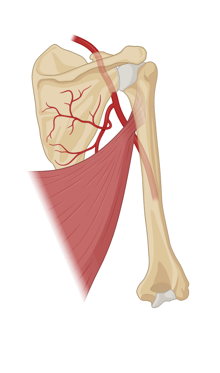
- Subscapularis – Origin: The subscapularisMedial rotator Front of shoulder blade; rotates arm inward. muscle originates from the subscapular fossaBroad, slightly concave surface on the anterior scapula that faces the ribcage., a concave depression on the anterior surface of the scapula (shoulder blade). It covers a significant portion of the scapula’s anterior surface. Insertion: The muscle fibers of the subscapularis converge to form a tendon. This tendon inserts onto the lesser tubercleSmaller bump anterior to head; also for muscle attachment. of the humerus. The lesser tubercle is a bony prominence on the front of the upper arm bone (humerus). Actions: Medial Rotation of the Shoulder Stabilization of the Shoulder Joint Contribution to Arm Abduction.
- Supraspinatus – Origin: The supraspinatusAbductor Top of shoulder blade; starts arm lift away from body. muscle originates from the supraspinous fossaShallow depression above the spine for muscle attachment.. This is a concavity in the scapula’s posterior surface. It is located above the spine of the scapula. Insertion: It inserts into the greater tubercle of the humerus. This is a prominent bony projection on the lateral aspect of the upper arm bone. Actions: Shoulder Abduction Stabilization of the Shoulder Joint.
Upper Arm muscles
- Biceps Brachii – Origin: Long Head: The long head of the biceps originates from the supraglenoid tubercle of the scapula. Short Head: The short head of the biceps originates from the coracoid process of the scapula. Lateral Head: The lateral head originates from the posterior surface of the humerus, above the radial groove. Insertion: The two heads of the biceps merge to form a single muscle belly. This muscle belly inserts onto the radial tuberosityRough bump just below head for biceps muscle attachment.. The radial tuberosity is a bony protuberance on the radiusLateral forearm bone (thumb side); rotates around ulna during pronation/supination. bone in the forearm. Additionally, the biceps brachiiFlexor / Supinator Front of upper arm; bends elbow and turns palm upward. inserts into the deep fascia of the forearm via the bicipital aponeurosis. Actions: Assistance in Shoulder Flexion Supination of the Forearm Elbow Flexion.
- Brachialis – Origin: The brachialisFlexor Under biceps; main muscle for elbow flexion muscle originates from the anterior surface of the humerus, specifically the distal half of the humerus. It arises along the entire length of the humerus. It starts just below the deltoid muscle’s insertion and extends down to the level of the elbow joint. Insertion: The brachialis muscle inserts onto the ulna, a bone in the forearm. More specifically, it attaches to the ulnar tuberosity and the coronoid process of the ulna. This attachment provides stability and support to the elbow joint. Actions: Stabilization of the Elbow Joint Support for the Biceps Brachii.
- Brachioradialis – Origin: The brachioradialisFlexor Forearm near elbow; helps flex arm in handshake position. muscle originates from the lateral supracondylar ridge of the humerus. This is a bony ridge on the lateral side of the lower end of the humerus. Insertion: The brachioradialis inserts onto the lateral surface of the distal end of the radius, near the styloid processThere are many styloid processes in the human body. All are pointed projections that serve as muscle. This specific insertion allows for its characteristic movements and functions. Actions: Elbow Flexion Forearm Supination and Pronation Stabilization during Elbow Movements.
- Triceps Brachii – Origin: Long Head: The long head of the triceps originates from the infraglenoid tubercle of the scapula. Lateral Head: The lateral head originates from the posterior surface of the humerus, above the radial groove. Medial Head: The medial head originates from the posterior surface of the humerus, below the radial groove. Insertion: The triceps brachiiExtensor Back of upper arm; straightens the elbow muscles merge into a single tendon. This tendon inserts into the olecranon processLarge, curved posterior projection—forms the bony point of the elbow. of the ulna. It forms the posterior border of the elbow joint. Actions: Elbow Extension Stabilization of the Elbow Joint Assistance in Shoulder Extension.
Identify More Muscles
Link to more Muscle Identification
Upper Arm Muscles
Forearm Muscles
Calf Muscles
Anterior Thigh Muscles
Posterior Thigh Muscles
Knee Structures
Chest Muscles
Back Muscles
Abdominal Muscles
Neck Muscles
Head Muscles
List of terms
- pubic symphysis
- pubic crest
- xiphoid process
- sternum
- ribs
- iliac crest
- anterior
- lateral
- lumbar vertebrae
- pubis
- adductor longus
- superior border
- linea aspera
- femur
- medial
- tibia
- proximal
- iliacus
- iliac fossa
- ilium
- lesser trochanter
- greater trochanter
- psoas major
- anterior inferior iliac spine
- acetabulum
- rectus femoris
- quadriceps tendon
- patella
- tibial tuberosity
- sartorius
- anterior superior iliac spine
- medial condyle
- superior
- spine
- lateral condyle
- vastus lateralis
- vastus medialis
- latissimus dorsi
- humerus
- rhomboid major
- thoracic vertebrae
- medial border
- scapula
- inferior angle
- rhomboid minor
- spinous process
- inferior
- teres major
- lateral border
- teres minor
- greater tubercle
- extensor digitorum longus
- fibula
- extensor digitorum
- tarsals
- phalanges
- head
- fibularis longus
- medial cuneiform
- lateral malleolus
- gastrocnemius
- calcaneus
- movement
- soleus
- deep
- tibialis anterior
- clavicle
- pectoralis minor
- cartilage
- coracoid process
- serratus anterior
- superior angle
- base
- dorsal
- extensor carpi ulnaris
- ulna
- pisiform
- hamate
- buccinator
- sphenoid
- mandible
- orbicularis oris
- mouth
- frontalis
- occipitalis
- skin
- superficial
- masseter
- zygomatic
- primary muscles
- mentalis
- occipital
- temporal bone
- lacrimal
- orbicularis oculi
- maxilla
- temporalis
- coronoid process
- zygomaticus major
- zygomaticus minor
- cervical vertebrae
- neck
- inspiration
- levator scapulae
- anterior scalene
- splenius capitis
- mastoid process
- sternocleidomastoid
- manubrium
- acromion process
- biceps femoris
- ischial tuberosity
- ischium
- gluteus maximus
- sacrum
- coccyx
- gluteus medius
- semimembranosus
- semitendinosus
- deltoid
- infraspinatus
- infraspinous fossa
- subscapularis
- subscapular fossa
- lesser tubercle
- supraspinatus
- supraspinous fossa
- radial tuberosity
- radius
- biceps brachii
- brachialis
- brachioradialis
- styloid process
- triceps brachii
- olecranon process