Time To Read
Date Last Modified
The anterior cruciate ligamentStabilizer Inside the knee; prevents shin bone from sliding too far forward (ACL) connects the femurThigh bone; longest and strongest bone in the body; has a large round head and prominent trochanters (thigh bone) to the tibiaShinbone; large, weight-bearing medial bone of the lower leg. (shin bone). It attaches from the posterior part of the inner surface of the lateralAway from the midline of the body. femoral condyle. It extends to the anteriorThe front of the body or toward the front when standing in the anatomical position. intercondylar area of the tibia. The ACL runs diagonally through the center of the knee. It crosses in front of the posterior cruciate ligamentStabilizer Inside knee; prevents shin from sliding backward. (PCL). This forms a crisscross shape—hence the name “cruciate.”
The ACL prevents the tibia from sliding too far forward relative to the femur, known as anterior translationThe process of converting mRNA into a protein.. It resists excessive rotational movements. This is crucial during activities like pivoting, jumping, and sudden stops or changes in direction. It also contributes to limiting hyperextension of the knee. Injury to the ACL is common in athletes. It can significantly impair movementA fundamental property of life involving motion of the body or its parts. and joint stability. Often, surgery and physical therapy are required for recovery.
Interactive Materials
🩻 Label or Draw on the Knee
Use the tools below to draw or add text labels directly on the pelvic girdle image. Choose a color, undo/redo strokes, clear your work, or save it when done.
Identify More Muscles
Link to more Muscle Identification
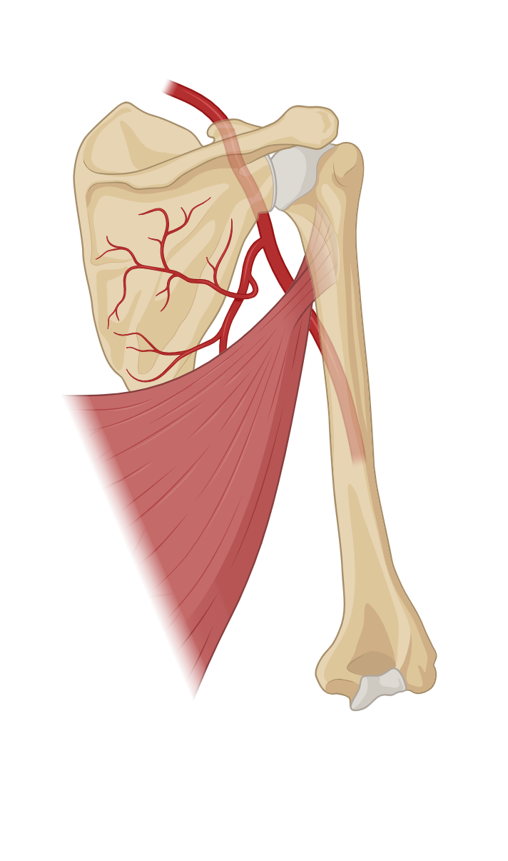
Upper Arm Muscles
Forearm Muscles
Calf Muscles
Anterior Thigh Muscles
Posterior Thigh Muscles
Knee Structures
Chest Muscles
Back Muscles
Abdominal Muscles
Neck Muscles
Head Muscles
List of terms
- anterior cruciate ligament
- femur
- tibia
- lateral
- anterior
- posterior cruciate ligament
- translation
- movement