Time To Read
Date Last Modified
The posterior cruciate ligamentStabilizer Inside knee; prevents shin from sliding backward. (PCL) is one of the two major internal stabilizing ligaments of the knee joint. It forms an X-shape with the anterior cruciate ligamentStabilizer Inside the knee; prevents shin bone from sliding too far forward (ACL). It starts at the anterolateral surface of the medialToward the midline of the body femoral condyle. Then, it inserts on the posterior aspect of the tibial intercondylar area. It is thicker and stronger than the ACL, making it less prone to injury.
The PCL’s primary role is to prevent the tibiaShinbone; large, weight-bearing medial bone of the lower leg. from sliding too far backward relative to the femurThigh bone; longest and strongest bone in the body; has a large round head and prominent trochanters. This prevents posterior translationThe process of converting mRNA into a protein., especially when the knee is bent. It also provides rotational stability. It helps resist forces that push the tibia backward during sudden stops. It also resists forces when falling onto a bent knee. While PCL injuries are less common than ACL injuries, they can occur in car accidents (“dashboard injuries”). They can also occur during sports. These injuries often require physical therapy. In severe cases, surgical repair may be needed to restore full
Interactive Materials
🩻 Label or Draw on the Knee
Use the tools below to draw or add text labels directly on the pelvic girdle image. Choose a color, undo/redo strokes, clear your work, or save it when done.
Identify More Muscles
Link to more Muscle Identification
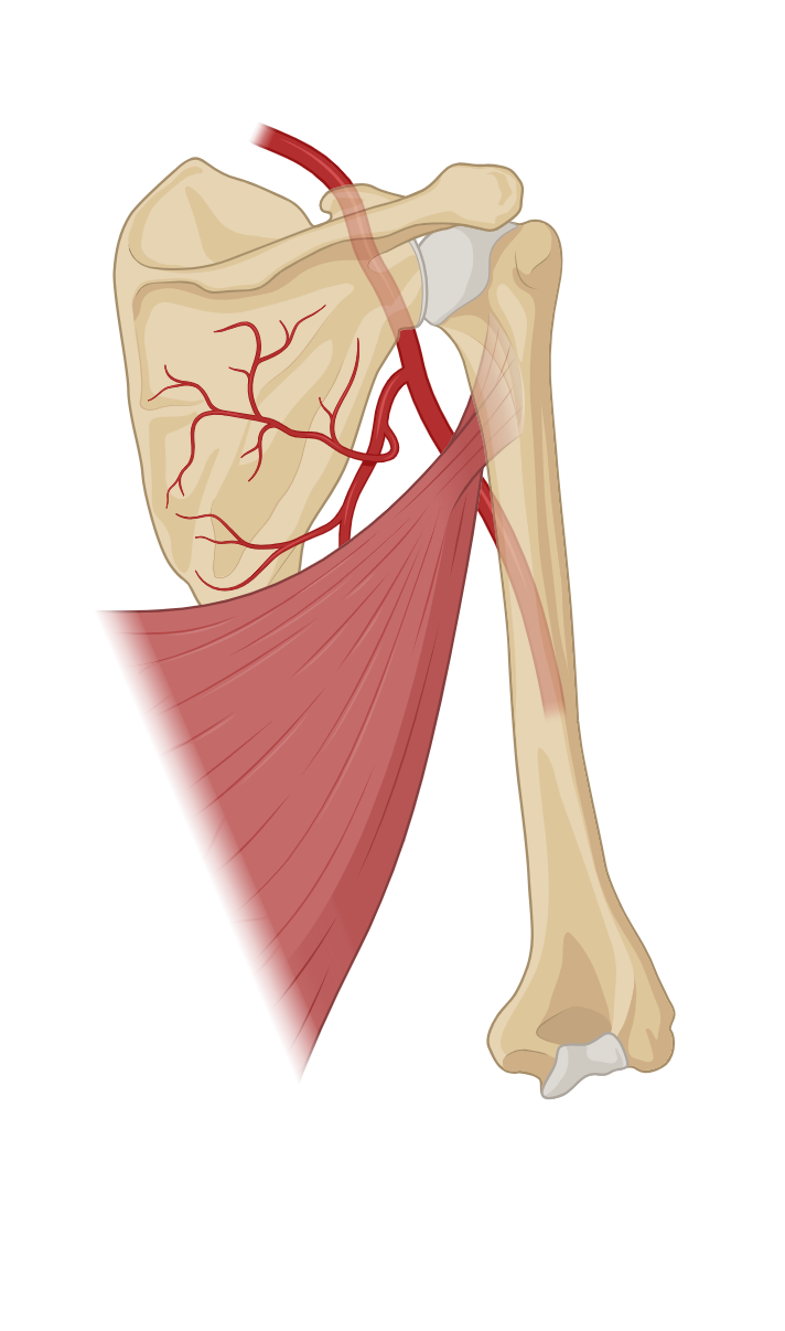
Upper Arm Muscles
Forearm Muscles
Calf Muscles
Anterior Thigh Muscles
Posterior Thigh Muscles
Knee Structures
Chest Muscles
Back Muscles
Abdominal Muscles
Neck Muscles
Head Muscles
List of terms
- posterior cruciate ligament
- anterior cruciate ligament
- medial
- tibia
- femur
- translation