Time To Read
Date Last Modified
The lateral collateral ligamentStabilizer Outer knee; resists outward bending. (LCL) connects the lateralAway from the midline of the body. epicondyle of the femurThigh bone; longest and strongest bone in the body; has a large round head and prominent trochanters. It attaches to the outer thigh bone. The LCL also connects to the headRounded proximal end that fits into the acetabulum of the hip bone. of the fibulaSlender lateral leg bone; stabilizes ankle but bears little weight.. The fibula is the smaller bone of the lower leg located just below the knee. Unlike the ACL and PCL, the LCL is not part of the joint capsule. It lies outside the synovial cavity. This makes it an extracapsular ligament.
The LCL’s primary role is to resist varus stress. This stress occurs when the knee is pushed outward. It moves away from the body’s midline. It also helps stabilize the knee during side-to-side movements, particularly preventing excessive side-bending or bowing of the knee. LCL injuries are less common than ACL injuries. However, they can occur with trauma or force. This usually happens when force is applied to the inner side of the knee. These injuries are especially common during sports or accidents involving sudden impacts.
Interactive Materials
🩻 Label or Draw on the Knee
Use the tools below to draw or add text labels directly on the pelvic girdle image. Choose a color, undo/redo strokes, clear your work, or save it when done.
Identify More Muscles
Link to more Muscle Identification
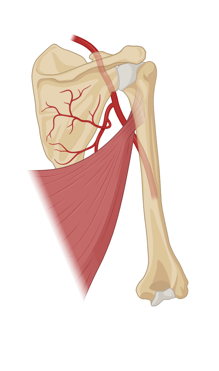
Upper Arm Muscles
Forearm Muscles
Calf Muscles
Anterior Thigh Muscles
Posterior Thigh Muscles
Knee Structures
Chest Muscles
Back Muscles
Abdominal Muscles
Neck Muscles
Head Muscles
List of terms
- lateral collateral ligament
- lateral
- femur
- head
- fibula