Time To Read
Date Last Modified
Pl. II · FAM. SIMPLICIDAE
Simplex cubicus var. vombati
the cube-maker · simple cuboidal epithelium
The plate
The specimen, drawn
Pl. II
The tissue as you will meet it on the slide.


Habitat & range
Where it lives
Body map
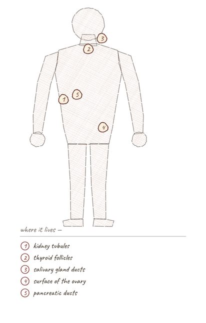
Where this tissue is found.
Range
- Kidney tubules
- Ducts of salivary, sweat and pancreatic glands
- Thyroid folliclesStructures in the ovaries that contain developing oocytes.
- Surface of the ovary
Collection record
- Add your slide numbers here.
Niche
What it does for a living
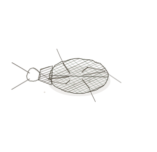
Taking in and handing back. Absorption and secretionThe process of moving substances from the blood into the nephron tubule to be excreted in urine. in small volumes, with enough cytoplasmThe gel-like substance within a cell that contains organelles and cytosol. to run the machinery but not enough height to slow traffic. This is the body’s standard-issue working lining.
field collected specimen
How to know it on sight
- One row of boxes, as tall as they are wide.
- A round nucleusThe control center of the cell that contains DNA and directs cellular activities. sitting in the middle of each.
- In a tubule cut crossways they ring the lumenThe inside space of a hollow organ or structure. like beads.
- Often brush-bordered on the apical surface in the kidney.
Specimen 1

Specimen 2

Specimen 3


People keep calling her by the wrong name
Look-alikes
Three plates. Read the line underneath each one before you decide.
Simple columnar

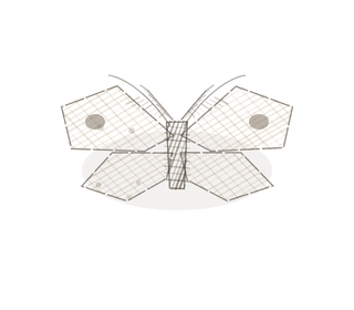
Tell them apart by height against width, and where the nucleus sits — centred in cuboidal, low in columnar.
Transitional, relaxed

Tell them apart by counting layers and looking at the top row. Transitional has several layers and a domed surface.
Stratified cuboidal

Tell them apart by the count. One row or two — that is the whole question.
Examine it yourself
The virtual slide
Zoom in until you can see the surface of the cellsThe basic structural and functional units of life., then find the basement membrane underneath.
Virtual slide courtesy of Histology Guide.
YOur missions
Field Notes for Future Missions
Mission 1: label the lining
H5P image hotspots — paste the shortcode here.
Mission 2: quick check
Field notes
Why this animal
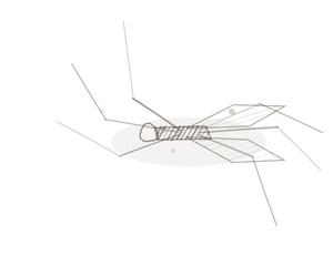
The wombat produces cube-shaped droppings — the only animal that does. Nothing else makes cubes on purpose, and nothing else makes “cuboidal” stick.
Next · Pl. III
Simplex columnaris var. penicillatus
the brush-bearer · simple columnar epithelium.
Hop to:
← Epithelial Tissues: A Guided Journey
All Modules
- Anatomical Language, Membranes & Homeostasis
- Just Enough Chemistry
- The Cell & Its Transport
- Making Cells & Proteins
- The Integumentary System
- The Skeletal System
- The Muscular System
- Nervous Tissue & the Senses
- The Spinal Cord
- The Brain & the Blood–Brain Barrier
- The Autonomic Nervous System
- Special Senses (in developmentThe process of growth and differentiation.)
- The Endocrine System
- Blood
- The Heart
- Blood Vessels
- The Digestive System
- The Respiratory System
- The Urinary System
- Fluids, Electrolytes & Acid–Base Balance
- The Reproductive System
- The Immune System
Plates and body maps drawn for this course. Micrographs from the BIO 171 slide collection. Virtual slides courtesy of Histology Guide.
List of terms
- follicles
- secretion
- cytoplasm
- nucleus
- lumen
- cells
- development