Time To Read
Date Last Modified
Pl. III · FAM. LAEVIDAE
Laevis fusiformis var. strangulans
the slow squeeze · smooth muscle
The plate
The specimen, drawn
Pl. I
[ plate drawing to come · the strangler fig ]

Habitat & range
Where it lives
Body map
[ body map to come — needs the BioRender export ]
Range
- The muscularis externaA smooth muscle layer of the digestive tract responsible for peristalsis and segmentation. of the whole gut, from the middle of the esophagusThe muscular tube that transports food from the pharynx to the stomach via peristalsis. to the anusThe terminal opening of the digestive tract through which feces are expelled..
- The wall of every artery and vein, wrapped circularly as the tunica mediaThe middle layer of a blood vessel, composed of smooth muscle and elastic tissue, responsible for va.
- The airways, all the way down to the terminal bronchiolesThe last portion of the conducting airway before respiratory bronchioles..
- The bladderA muscular organ that stores urine before excretion., the ureters, the uterusThe muscular organ where a fertilized egg implants and develops. and the ductus deferens.
- The iris and ciliary body of the eye — and the tiny muscles that stand your hair up.
Collection record
- Add your slide numbers here.
Niche
What it does for a living
Slow, cheap and tireless. Smooth muscle narrows tubes and empties hollow organs, and it does the work on a budget: it can hold a contraction for hours on a fraction of the ATPThe energy currency of cells used for muscle contraction. striated muscle would burn through, and it can be stretched enormously and still pull. It also never takes an order from you. Autonomic nerves, circulating hormones and the stretch of the organ wall itself all get a vote, and the sheet answers as one piece because the cellsThe basic structural and functional units of life. are wired to each other by gap junctions rather than each waiting for its own nerve.
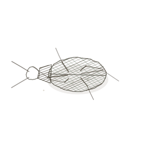
field collected specimen
How to know it on sight
- No striations anywhere — the cytoplasmThe gel-like substance within a cell that contains organelles and cytosol. is an even pink.
- Cells spindle-shaped: fat through the middle, tapering to a point at both ends.
- One central nucleusThe control center of the cell that contains DNA and directs cellular activities. to a cell, and in a good longitudinal section A cut or slice of the body or an organ for study. the nucleiClusters of neurons in the CNS responsible for processing information. look staggered, because the fat middles do not line up with each other.
- Sheets rather than bundles — usually two layers lying at right angles to one another.

Specimen 1

Ureter. The even pink with no cross-stripes anywhere is the diagnosis; the nuclei are inside the pink, one to a cell.
Specimen 2

Detrusor. Two layers meet in this field — one cut lengthwise into long spindles, one cut across into circles.
Specimen 3

Esophagus, cut across. Each cell is a small pink polygon, and only the ones sliced through the middle show you a nucleus at all.


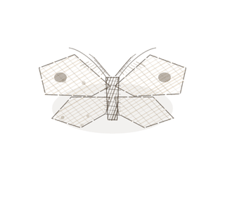
People keep calling her by the wrong name
Look-alikes
Two plates. Read the line underneath each one before you decide.
Dense regular connective tissue

Tell them apart by where the nuclei sit. Smooth muscle nuclei are inside the pink; tendon nuclei lie in rows in the gaps between the bundles.
Cardiac muscle

Tell them apart by the stripes and the fork. Cardiac is striated and branches; smooth is evenly pink and never branches.
Examine it yourself
The virtual slide
Find a patch where the cells are cut lengthwise and look for the tapered ends. Then find a patch cut across, and notice that only some of the cells show a nucleus.
Virtual slide courtesy of Histology Guide.
YOur missions
Field Notes for Future Missions
Mission 1: find the spindles
Mission 2: quick check
Field notes
Why this organism
A strangler fig begins as a seed dropped in the crown of another tree, sends roots down around its host, and over decades tightens until the tree inside it is gone. Nothing about that is fast and nothing about it is voluntary. Smooth muscle does the same thing to a tube: a slow, sustained, entirely unhurried squeeze that costs almost nothing to hold and that you cannot argue your way out of. It is why you cannot simply decide to stop a cramp.
Return · Ordo Musculi
Frontispiece
the three specimens, and the key that tells them apart.
Hop to:
← Epithelial Tissues: A Guided Journey
All plates — The Tissue Field Guide: Epithelium
Frontispiece Ordo Epithelia (main page)
Pl. I · Simple squamous
Pl. II · Simple cuboidal
Pl. III · Simple columnar
Pl. IV · Stratified squamous
Pl. V · Stratified cuboidal
Pl. VI · Stratified columnar
Pl. VII · Transitional
Pl. VIII · Pseudostratified columnar
Epithelial Identification Game
All Modules
- Anatomical Language, Membranes & Homeostasis
- Just Enough Chemistry
- The Cell & Its Transport
- Making Cells & Proteins
- The Integumentary System
- The Skeletal System
- The Muscular System
- Nervous Tissue & the Senses
- The Spinal Cord
- The Brain & the Blood–Brain Barrier
- The Autonomic Nervous System
- The Special Senses
- The Endocrine System
- Blood
- The Heart
- Blood Vessels
- The Digestive System
- The Respiratory System
- The Urinary System
- Fluids, Electrolytes & Acid–Base Balance
- The Reproductive System
- The Immune System
List of terms
- muscularis externa
- esophagus
- anus
- tunica media
- terminal bronchioles
- bladder
- uterus
- ATP
- cells
- cytoplasm
- nucleus
- section
- nuclei