Time To Read
Date Last Modified
Pl. II · FAM. PERIPHERICIDAE
Periphericus globosus var. allianus
the garlic bulb · a ganglion
The plate
The specimen, drawn
Pl. II

The tissue as you will meet it on the slide.

Habitat & range
Where it lives
Body map

Where this tissue is found.
Range
- Dorsal root ganglia — one pair on every segment of the cord, sitting just outside it.
- The sympathetic chain, a string of them running the whole length of the spineProminent ridge on the posterior scapula dividing it into supraspinous and infraspinous fossae..
- Scattered through the wall of the gut, running it without ever asking you.
- And several more in the headRounded proximal end that fits into the acetabulum of the hip bone., beside the cranial nervesNerves that arise from the brain and control head and neck functions..
Collection record
- Add your slide numbers here.
Niche
What it does for a living
Housing. A ganglion is not really a relay and mostly not a decision; it is where the cell bodies of peripheral neuronsThe functional cells of the nervous system that transmit signals. are kept, parked outside the central nervous systemComposed of the brain and spinal cord; integrates and processes information. while their axons run off somewhere else entirely. A dorsalRelating to the back side of the body. root ganglion holds the cell bodyThe central part of a neuron containing the nucleus and organelles. of every sensory neuron for its segment. The cell is a T-junction — one long process out to the skinThe body’s largest organ, providing protection and regulation., another into the cord — and the cell body itself sits off to one side doing the housekeeping that keeps both of them alive.
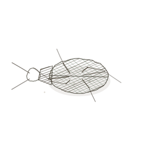
field collected specimen
How to know it on sight
- Large, round, pale cell bodies — among the biggest cellsThe basic structural and functional units of life. you will meet on any slide.
- A nucleusThe control center of the cell that contains DNA and directs cellular activities. in the middle of each, with one obvious dark nucleolus in it.
- A ring of small dark satellite cell nucleiClusters of neurons in the CNS responsible for processing information. hugging the outside of every cell body.
- The whole crowd packed inside a capsule, usually with a nerve running out of one end.

Specimen 1

Low power. The ganglion is the swelling beside the cord — same segment, different job.
Specimen 2

The cell bodies at higher power. Look for the single dark nucleolus in each pale nucleus.
Specimen 3

A parasympathetic ganglion in the wall of an organ — smaller cells, same plan, same wrappers.


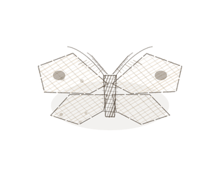
People keep calling her by the wrong name
Look-alikes
Two plates. Read the line underneath each one before you decide.
Peripheral nerve

Tell them apart by what is there. A nerve is axons and sheaths with no cell bodies at all; a ganglion is cell bodies, and the nerve is what leaves it.
Grey matter of the spinal cord

Tell them apart by the ring. Cord neurons sit in a tangle of fibres with glia between them; ganglion cells each wear a tidy ring of satellite nuclei.
Examine it yourself
The virtual slide
Find one cell body and go all the way round its edge, looking for the ring of small dark nuclei. Then check that no two cell bodies are touching.
Virtual slide courtesy of Histology Guide.
YOur missions
Field Notes for Future Missions
Mission 1: find the wrapper
Mission 2: quick check
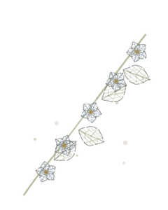
Field notes
Why this organism
A garlic bulb is one head holding a dozen bodies, and the thing nobody notices is that every clove has a papery wrapper of its own inside the shared skin. A ganglion is built on exactly that plan. The capsule is the outer skin, the neuron cell bodies are the cloves, and the ring of satellite cells around each one is a wrapper belonging to that cell alone. No cell body touches its neighbour. Once you have seen the ring you will not mistake a ganglion for anything else again.
Next · Pl. III
Centralis trilaminatus var. thujanus
the tree of life · cerebellumThe brain region responsible for balance, coordination, and fine motor control..
Hop to:
← Epithelial Tissues: A Guided Journey
All plates — The Tissue Field Guide: Epithelium
Frontispiece Ordo Epithelia (main page)
Pl. I · Simple squamous
Pl. II · Simple cuboidal
Pl. III · Simple columnar
Pl. IV · Stratified squamous
Pl. V · Stratified cuboidal
Pl. VI · Stratified columnar
Pl. VII · Transitional
Pl. VIII · Pseudostratified columnar
Epithelial Identification Game
All Modules
- Anatomical Language, Membranes & Homeostasis
- Just Enough Chemistry
- The Cell & Its Transport
- Making Cells & Proteins
- The Integumentary System
- The Skeletal System
- The Muscular System
- Nervous Tissue & the Senses
- The Spinal Cord
- The Brain & the Blood–Brain Barrier
- The Autonomic Nervous System
- The Special Senses
- The Endocrine System
- Blood
- The Heart
- Blood Vessels
- The Digestive System
- The Respiratory System
- The Urinary System
- Fluids, Electrolytes & Acid–Base Balance
- The Reproductive System
- The Immune System
List of terms
- ganglion
- spine
- head
- cranial nerves
- neurons
- central nervous system
- dorsal
- cell body
- skin
- cells
- nucleus
- nuclei
- cerebellum