Time To Read
Date Last Modified
Pl. V · FAM. CENTRALIDAE
Centralis inversus var. interrogationis
the butterfly · the spinal cord
The plate
The specimen, drawn
Pl. V

The tissue as you will meet it on the slide.

Habitat & range
Where it lives
Body map

Where this tissue is found.
Range
- From the baseA substance that accepts hydrogen ions (H⁺) or releases hydroxide ions (OH⁻). of the skull down to about the first lumbar vertebra — shorter than the spineProminent ridge on the posterior scapula dividing it into supraspinous and infraspinous fossae. it lives in.
- Two swellings along its length: one wiring the arms, one wiring the legs.
- Below where it ends, a spray of roots and no cord at all.
Collection record
- Add your slide numbers here.
Niche
What it does for a living
A two-way trunk road with its own local government. Everything going up to the brain and everything coming down from it passes through the cord in named tractsBundles of nerve fibers in the CNS that carry signals between brain regions. in the white matterThe outer portion of the spinal cord made of myelinated nerve fibers that transmit signals.. But the cord is not only wiring. The grey matter in the middle holds the cell bodies that fire muscles directly, and a reflex can start and finish there without the brain being consulted at all — which is why your hand is off the hot pan before you have felt anything.
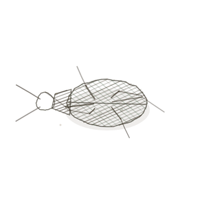
field collected specimen
How to know it on sight
- Grey on the inside, white on the outside — the exact reverse of the cerebrumThe largest part of the brain, responsible for thought, memory, and voluntary movements..
- The grey is a butterfly, or an H: narrow dorsalRelating to the back side of the body. horns, blunt ventralRelating to the front or belly side of the body. horns.
- A tiny central canalA hollow canal in an osteon containing blood vessels and nerves. sitting in the middle of the crossbar.
- A deepAway from the surface of the body. fissureA deep groove in the brain’s surface. at the front and a shallow septum at the back, which is how you tell which way is up.

Specimen 1

Whole cord at low power, with a ganglionA cluster of neuron cell bodies located in the peripheral nervous system (PNS). beside it. The butterfly is the diagnosis.
Specimen 2

The same arrangement in Masson. Different stain, identical shape — the shape is what you are learning.
Specimen 3

The horns are inhabited: different jobs live in different corners of the grey.


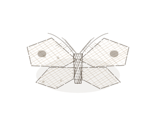
People keep calling her by the wrong name
Look-alikes
Two plates. Read the line underneath each one before you decide.
Cerebrum

This is the one that matters. Both are central nervous tissue; the cerebrum keeps its grey outside, the cord keeps its grey inside.
Dorsal root ganglion

They share a slide, so read position. The ganglion is the bump off to one side; the cord is the round thing with the butterfly in it.
Examine it yourself
The virtual slide
Find the deep fissure first and put it at the bottom. Then name the narrow horns and the blunt ones, and look for the canal in the crossbar.
Virtual slide courtesy of Histology Guide.
YOur missions
Field Notes for Future Missions
Mission 1: which way is up
Mission 2: quick check
Field notes
Why this organism
Polygonia interrogationis is named for a small silver mark on the underside of its hindwing, shaped like a question mark. But the thing that identifies it in the field is not the mark — it is the outline. The wing margins are ragged and angular, never a smooth curve, and nothing else in the northeast is shaped quite like it. The spinal cord is the same kind of problem. You do not identify it by colour or by stain; you identify it by a shape, grey horns sitting in the middle of a pale rim. Once you can see the butterfly you have the answer before you have looked at anything else on the slide.
Next · Pl. VI
Centralis trimembranatus var. arachnoideus
the three veils · the meninges(singular: meninx) – Protective membranes surrounding the spinal cord and brain..
Hop to:
← Epithelial Tissues: A Guided Journey
All plates — The Tissue Field Guide: Epithelium
Frontispiece Ordo Epithelia (main page)
Pl. I · Simple squamous
Pl. II · Simple cuboidal
Pl. III · Simple columnar
Pl. IV · Stratified squamous
Pl. V · Stratified cuboidal
Pl. VI · Stratified columnar
Pl. VII · Transitional
Pl. VIII · Pseudostratified columnar
Epithelial Identification Game
All Modules
- Anatomical Language, Membranes & Homeostasis
- Just Enough Chemistry
- The Cell & Its Transport
- Making Cells & Proteins
- The Integumentary System
- The Skeletal System
- The Muscular System
- Nervous Tissue & the Senses
- The Spinal Cord
- The Brain & the Blood–Brain Barrier
- The Autonomic Nervous System
- The Special Senses
- The Endocrine System
- Blood
- The Heart
- Blood Vessels
- The Digestive System
- The Respiratory System
- The Urinary System
- Fluids, Electrolytes & Acid–Base Balance
- The Reproductive System
- The Immune System
List of terms
- spinal cord
- base
- spine
- tracts
- white matter
- cerebrum
- dorsal
- ventral
- central canal
- deep
- fissure
- ganglion
- meninges