Time To Read
Date Last Modified
Pl. III · FAM. LAXIDAE
Laxus reticularis var. mycelialis
the invisible net · reticular connective tissue
The plate
The specimen, drawn
Pl. III

A fine branching mesh of type III collagenA structural protein in the dermis that provides strength and elasticity., holding soft organs open from the inside. You will not see it in H&E — it takes silver.

Habitat & range
Where it lives
Body map

Wherever blood cellsThe basic structural and functional units of life. or immune cells need somewhere to sit, there is a reticular net holding the room open for them.
Range
- Lymph nodes, in chains through the neckNarrow region just below the head; common fracture site., armpit, chest, abdomen and groin.
- The spleen, as the framework of both red and white pulp.
- Red bone marrow, holding the developing blood cells in place.
- The liverA large organ that produces bile, detoxifies blood, and stores nutrients., as the scaffold the hepatocyte plates are built on.
- Anywhere soft tissue needs a skeleton that does not take up room.
Collection record
- Add your slide numbers here.
Niche
What it does for a living
Being the furniture. Reticular tissue makes a three-dimensional scaffold out of fine branching type III collagen, and the cells that matter — lymphocytes, macrophages, developing blood cells — live in its spaces rather than being part of it. It holds a soft organ open without making it stiff, which is why you can squeeze a lymph node and it springs back.
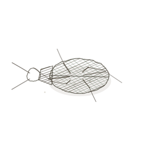
field collected specimen
How to know it on sight
- A fine black or dark brown branching net — silver stain, not H&E.
- Fibres are thin, even in calibre, and they branch and rejoin rather than running in bundles.
- The spaces are full of free cells that are not attached to the fibres.
- No single direction; the net is three-dimensional and looks tangled in any one plane.

Specimen 1

A lymph node at high power. Follow one fibre to where it branches, and then follow both branches.
Specimen 2

Marrow. Here the net holds developing blood cells and fat cells together in the same space.

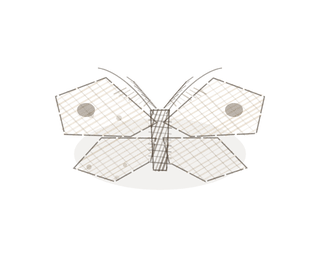
People keep calling her by the wrong name
Look-alikes
Two plates. Read the line underneath each one before you decide.
Areolar connective tissue

Areolar also has no grain, but its fibres are thick pink bundles of type I; reticular fibres are fine, even, and only show up with silver.
Elastic connective tissue

Verhoeff makes elastin dark too. Elastic fibres run in long wavy parallel lines; reticular fibres branch into a net and go nowhere in particular.
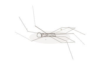
Examine it yourself
The virtual slide
Zoom in on a lymphoid follicle until the individual lymphocytes separate. The net holding them is reticular tissue, and in this stain you will have to infer it from the spaces rather than see it directly.
Virtual slide courtesy of Histology Guide.
YOur missions
Field Notes for Future Missions
Mission 1: follow the net
Mission 2: quick check
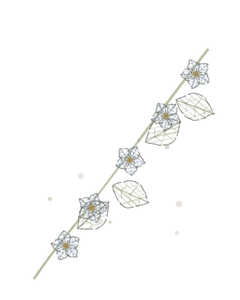
Field notes
Why this organism
A mycelium is the largest part of a fungus and the part nobody sees. It threads through the whole of the soil as a branching net, holding the ground together and feeding everything that lives in it, and then puts up a mushroom for a week. Reticular tissue works the same way round: the net is the organ, and the cells you actually came to look at are just the things living in it.
Next · Pl. IV
Densus rectus var. lineus
captain lengthwise · dense regular connective tissue.
Hop to:
← Epithelial Tissues: A Guided Journey
All plates — The Tissue Field Guide: Epithelium
Frontispiece Ordo Epithelia (main page)
Pl. I · Simple squamous
Pl. II · Simple cuboidal
Pl. III · Simple columnar
Pl. IV · Stratified squamous
Pl. V · Stratified cuboidal
Pl. VI · Stratified columnar
Pl. VII · Transitional
Pl. VIII · Pseudostratified columnar
Epithelial Identification Game
All Modules
- Anatomical Language, Membranes & Homeostasis
- Just Enough Chemistry
- The Cell & Its Transport
- Making Cells & Proteins
- The Integumentary System
- The Skeletal System
- The Muscular System
- Nervous Tissue & the Senses
- The Spinal Cord
- The Brain & the Blood–Brain Barrier
- The Autonomic Nervous System
- The Special Senses
- The Endocrine System
- Blood
- The Heart
- Blood Vessels
- The Digestive System
- The Respiratory System
- The Urinary System
- Fluids, Electrolytes & Acid–Base Balance
- The Reproductive System
- The Immune System
List of terms
- collagen
- cells
- neck
- liver