Time To Read
Date Last Modified
Pl. VII · FAM. SUSTINENTIDAE
Sustinens hyalinus var. tremellus
the glider · hyaline cartilageA flexible connective tissue found in joints, the ear, nose, and rib cage. Cartilage can be of three
The plate
The specimen, drawn
Pl. VII

A matrix so fine-grained it looks like frosted glass. The fibres are there; they are just too small and too close in refractive index to see.

Habitat & range
Where it lives
Body map

Hyaline is the commonest cartilage in the body, and it goes wherever something has to slide or stay open.
Range
- The articular surface of every synovial joint.
- The rings of the trachea and the plates of the bronchiThe large airways that branch from the trachea into the lungs, dividing into smaller bronchioles..
- Where each rib meets the sternumFlat bone forming the center of the chest; anchor for ribs and clavicles. Appears as a vertical dagg, as costal cartilage.
- The end of the nose, and most of the larynxThe voice box; contains vocal cords and connects the pharynx to the trachea..
- The whole fetal skeleton, before bone replaces it.
Collection record
- Add your slide numbers here.
Niche
What it does for a living
Being slippery and staying open. Articular hyaline is the lowest-friction surface in biology, and it does that with a smooth matrix and a great deal of bound waterThe universal solvent essential for life.. It is avascularTo be devoid of blood capillaries. Epithelial tissue is avascular, kind of like a cap of dead cells — chondrocytes are fed by diffusionPassive movement of molecules from areas of high to low concentration. through the matrix — which is why cartilage heals so badly, and why a torn knee surface does not simply grow back.
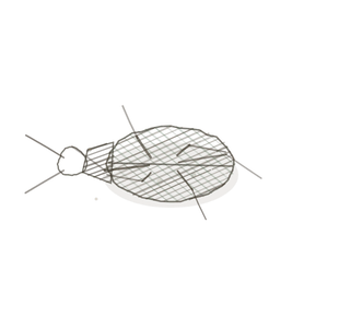
field collected specimen
How to know it on sight
- A smooth glassy matrix with no visible fibres at all.
- ChondrocytesMature cartilage cells housed in lacunae. sitting in lacunae — little holes with a cell in each.
- Cells often in pairs or small groups, where one divided and the daughters stayed put.
- A perichondrium at the edge, unless the surface is articular, in which case there is none.


Specimen 2

Trachea at low power. The cartilage is the deepAway from the surface of the body. block; follow it to its edge and find the perichondrium.
Specimen 3

Masson’s trichrome. A different stain, the same glassy matrix and the same lacunae.

People keep calling her by the wrong name
Look-alikes
Two plates. Read the line underneath each one before you decide.
Elastic cartilage

Both have chondrocytes in lacunae. Hyaline matrix is featureless; elastic matrix has a dark fibre network running through it.
Fibrocartilage

Fibrocartilage has obvious thick collagenA structural protein in the dermis that provides strength and elasticity. bundles between its cells. If you can see bundles, it is not hyaline.
Examine it yourself
The virtual slide
Find the tracheal cartilage and travel from its middle out to its edge. The cells get flatter and smaller as you approach the perichondrium, because that is where new cartilage is added.
Virtual slide courtesy of Histology Guide.
YOur missions
Field Notes for Future Missions
Mission 1: find a lacuna
Mission 2: quick check
Field notes
Why this organism
Snow fungus is a cluster of translucent gelatinous lobes that looks like it should not hold its own weight and does. There is structure in it, but you cannot see the structure — it is dispersed through a clear gel, which is the whole trick. Hyaline cartilage is exactly that: collagen II is in there in quantity, but the fibrils are thin and the matrix around them bends light the same way, so the tissue reads as frosted glass with cells in it.
Next · Pl. VIII
Sustinens elasticus var. auritus
the bouncer · elastic cartilageA flexible cartilage type found in the ear and epiglottis..
Hop to:
← Epithelial Tissues: A Guided Journey
All plates — The Tissue Field Guide: Epithelium
Frontispiece Ordo Epithelia (main page)
Pl. I · Simple squamous
Pl. II · Simple cuboidal
Pl. III · Simple columnar
Pl. IV · Stratified squamous
Pl. V · Stratified cuboidal
Pl. VI · Stratified columnar
Pl. VII · Transitional
Pl. VIII · Pseudostratified columnar
Epithelial Identification Game
All Modules
- Anatomical Language, Membranes & Homeostasis
- Just Enough Chemistry
- The Cell & Its Transport
- Making Cells & Proteins
- The Integumentary System
- The Skeletal System
- The Muscular System
- Nervous Tissue & the Senses
- The Spinal Cord
- The Brain & the Blood–Brain Barrier
- The Autonomic Nervous System
- The Special Senses
- The Endocrine System
- Blood
- The Heart
- Blood Vessels
- The Digestive System
- The Respiratory System
- The Urinary System
- Fluids, Electrolytes & Acid–Base Balance
- The Reproductive System
- The Immune System
List of terms
- hyaline cartilage
- cartilage
- bronchi
- sternum
- larynx
- water
- avascular
- diffusion
- Chondrocytes
- lacuna
- cells
- deep
- collagen
- elastic cartilage