Time To Read
Date Last Modified
Pl. XI · FAM. SUSTINENTIDAE
Sustinens osseus var. trabecularis
the lattice · spongy bone
The plate
The specimen, drawn
Pl. XI

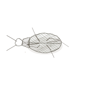
The same bone matrix, built as an open lattice of struts. Nearly the strength at a fraction of the weight — and the marrow lives in the gaps.

Habitat & range
Where it lives
Body map

Spongy bone fills the inside of flat bones and the ends of long ones, wherever the load arrives from several directions.
Range
- The body of every vertebra.
- The expanded ends of the long bones, under the joint surfaces.
- The blades of the pelvis.
- The sternumFlat bone forming the center of the chest; anchor for ribs and clavicles. Appears as a vertical dagg and the ribsCurved bones forming the rib cage; articulate with thoracic vertebrae and most with the sternum. — which is where marrow is sampled from.
- Between the two tables of the skull, as the diploe.
Collection record
- Add your slide numbers here.
Niche
What it does for a living
Carrying load at the lowest possible weight, and housing the marrow. The trabeculae are not random: they line up along the lines of stress that bone actually experiences, and they re-align if the stress changes. That is why weight-bearing exercise builds bone and why bed rest loses it, and why osteoporosis shows first as a thinning lattice rather than a thinning shell.
field collected specimen
How to know it on sight
- Irregular struts and plates of bone — trabeculae — with wide spaces between them.
- No osteonsThe structural unit of compact bone, consisting of concentric lamellae around a central canal.; the trabeculae are too thin to need central canalsA hollow canal in an osteon containing blood vessels and nerves..
- OsteocytesMature bone cells that maintain bone tissue. in lacunae within the struts, fed by diffusionPassive movement of molecules from areas of high to low concentration. from the marrow.
- The spaces are full of marrow, not empty, though processing often washes it out.

Specimen 1

Masson’s trichrome. The teal matrix is bone; the empty-looking spaces held marrow.

People keep calling her by the wrong name
Look-alikes
Two plates. Read the line underneath each one before you decide.
Compact bone

Compact is solid with concentric osteons; spongy is an open lattice with no rings at all.
Adipose tissue

Marrow in the spaces has fat cellsThe basic structural and functional units of life. in it, which can read as adipose — but adipose has no bony struts running through it.
Examine it yourself
The virtual slide
Both kinds on one slide. Find the boundary where the solid shell becomes the open lattice, and notice that it is a gradual change rather than a line.
Virtual slide courtesy of Histology Guide.
YOur missions
Field Notes for Future Missions
Mission 1: read the lattice
Mission 2: quick check
Field notes
Why this organism
A reef is not a solid wall. It is an open framework of branching struts with waterThe universal solvent essential for life. moving through every gap, and that is what lets it be enormous without being impossibly heavy — and what gives everything else somewhere to live. Spongy bone made the same decision. Solid bone throughout would be too heavy to carry, so the inside is built as a lattice aligned to the loads, and the marrow gets the spaces.
Next · Pl. XII
Sustinens transiens var. bambusinus
the shapeshifter · the epiphyseal plateA growth plate in developing bones made of cartilage..
Hop to:
← Epithelial Tissues: A Guided Journey
All plates — The Tissue Field Guide: Epithelium
Frontispiece Ordo Epithelia (main page)
Pl. I · Simple squamous
Pl. II · Simple cuboidal
Pl. III · Simple columnar
Pl. IV · Stratified squamous
Pl. V · Stratified cuboidal
Pl. VI · Stratified columnar
Pl. VII · Transitional
Pl. VIII · Pseudostratified columnar
Epithelial Identification Game
All Modules
- Anatomical Language, Membranes & Homeostasis
- Just Enough Chemistry
- The Cell & Its Transport
- Making Cells & Proteins
- The Integumentary System
- The Skeletal System
- The Muscular System
- Nervous Tissue & the Senses
- The Spinal Cord
- The Brain & the Blood–Brain Barrier
- The Autonomic Nervous System
- The Special Senses
- The Endocrine System
- Blood
- The Heart
- Blood Vessels
- The Digestive System
- The Respiratory System
- The Urinary System
- Fluids, Electrolytes & Acid–Base Balance
- The Reproductive System
- The Immune System
List of terms
- spongy bone
- sternum
- ribs
- osteons
- central canals
- Osteocytes
- diffusion
- cells
- water
- epiphyseal plate