Time To Read
Date Last Modified
Frontispiece · ORD. PNEUMATICA
Ordo Pneumatica
the air-movers · the respiratory tract
The plates
The five specimens
Three conducting airways, one place where gas actually crosses, and a lid. The key below covers the first four — a lid is not an airway and does not key like one.
Pl. I
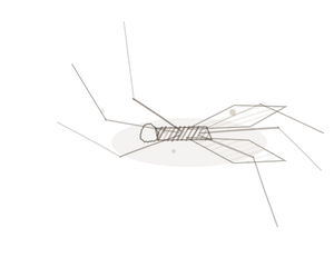
Anulatus ciliatus var. dasypus
the banded hose · trachea

Open Pl. I →
Pl. II
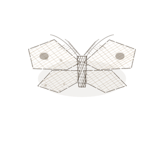
Lamellatus ciliatus var. mississippiensis
the plate armor · bronchus

Open Pl. II →
Pl. III
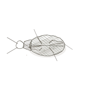
Nudus cuboidus var. actinius
the clencher · bronchiole

Open Pl. III →
Pl. IV
Sacculus squamosus var. riparius
the thin skinSkin found on most of the body, lacking a stratum lucidum. · alveolus

Open Pl. IV →
Pl. V
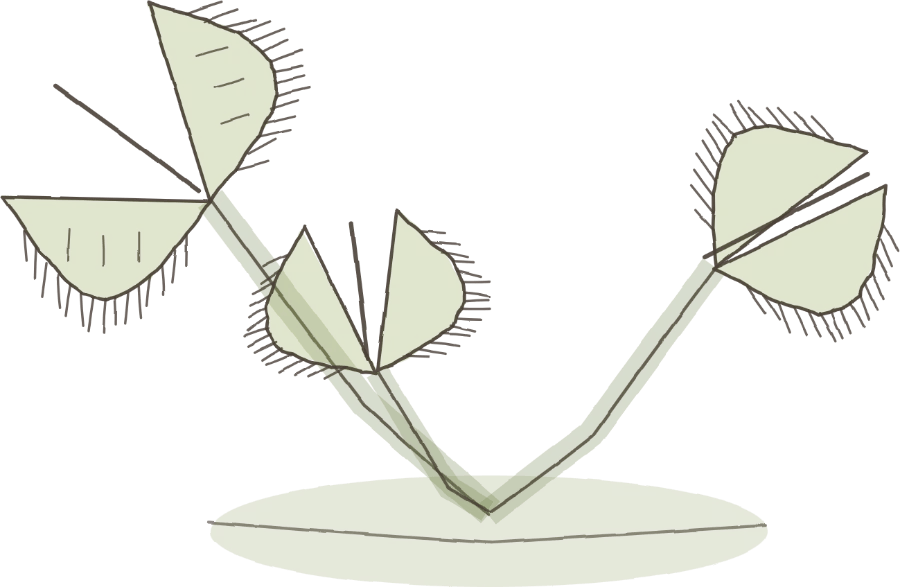
Operculum stratificatus var. dionaeus
the trapdoor · epiglottisA flap of cartilage that covers the trachea during swallowing to prevent food from entering the airw
Open Pl. V →
The ground plan
One tube, getting simpler
Everything from the nose down to the last terminal bronchiole only moves and conditions air. Not one molecule of oxygen crosses anywhere in it. The lung is a plumbing-and-air-conditioning system with a gas exchanger bolted on the end, and the exchanger is the last tenth of it.
Fig. 1 · one tube, getting simpler

Four steps, left to right — and only the last of them exchanges anything.
The five words
- Conducting zone — nose to terminal bronchiole. Moves and conditions air, and exchanges nothing.
- Respiratory zone — from the respiratory bronchiole onward. Anywhere an alveolus opens off the wall.
- Respiratory epithelium — the ciliated lining with goblet cellsThe basic structural and functional units of life., running from the nose down into the bronchiThe large airways that branch from the trachea into the lungs, dividing into smaller bronchioles..
- Lamina propria — the loose connective tissue under the lining, carrying the vessels and the glands.
- Adventitia — the outermost coat, binding the airway to whatever happens to be next to it.
Decide which zone you are in before anything else. It settles half the question on its own.

First question
What holds it open?
Go round the wall once and find the stiff part — if there is one at all.
Fig. 2 · what holds it open

Four walls, four answers. Only the first two have anything stiff in them.
- A ring — one continuous C of hyaline cartilageThe most abundant cartilage type, found in joints, ribs, and the nose., open at the back. Trachea.
- Plates — scattered islands of the same cartilageA flexible connective tissue found in joints, the ear, nose, and rib cage. Cartilage can be of three, with a complete muscle ring inside them. Bronchus.
- Nothing — muscle alone, and a lumenThe inside space of a hollow organ or structure. crimped into folds. Bronchiole.
- Not a tube at all — a cluster of thin-walled sacs. Alveolus.
The test is cartilage, and it is never size. A large bronchiole is wider than a small bronchus.
Second question
What is the lining doing?
Look at the cells on the free surface, and at what is sitting among them.
Fig. 3 · what the lining is doing

Tall and ciliated with goblet cells; shorter and ciliated with club cells; then one cell thick with nothing on it.
- Tall, pseudostratified, ciliated, with goblet cells — trachea and bronchus.
- Shorter, simple, ciliated, with club cells and no goblet cells — bronchiole.
- One cell thick with nothing on it, and a domed type II cell here and there — alveolus.
Goblet cells quit before the ciliaHair-like projections on the surface of some cells that move fluids or particles. do. Cilia with no goblet cell anywhere in the field means you are already past the bronchi — and that one factA statement based on direct observation that is repeatedly confirmed. will place you in the lung faster than anything else on this page.

The naming key
How to read a name
Put the two answers together: what holds it open first, then what the lining is doing.
First word · what holds it open
- Anulatus — held open by rings
- Lamellatus — held open by scattered plates
- Nudus — nothing holds it open; muscle alone
- Sacculus — not a tube at all, a sac
- Operculum — not a tube either; a lid
Second word · the lining
- ciliatus — pseudostratified, ciliated, goblet cells present
- cuboidus — simple ciliated cuboidal; club cells, no goblets
- squamosus — simple squamous, one cell thick
- stratificatus — stratified on one face, respiratory on the other
Worked both ways
- Nudus cuboidus → no cartilage, ciliated cuboidal, no goblet cells → a bronchiole.
- Anulatus ciliatus → a ring of cartilage with a tall ciliated lining → the trachea.
- You saw islands of cartilage and a ciliated lining with goblet cells, surrounded by lung → Lamellatus ciliatus, a bronchus.
Three that trip people up
The look-alikes
Three pairs get mixed up more than anything else in this volume. Two of them are below; the third — the epiglottis against the oesophagus — is worked through on Pl. V.
Fig. 4 · three that trip people up

Cartilage, then wall, then faces.
A small bronchus, or a large bronchiole?
The trap: sorting by diameter.
A large bronchiole is genuinely wider than a small bronchus, so size will mislead you every single time.
Hunt for cartilage, and hunt properly — one small island of it is enough to make the thing a bronchus.
A respiratory bronchiole, or an alveolar duct?
The trap: both have alveoliMicroscopic air sacs in the lungs where gas exchange occurs between air and blood. opening off them.
A respiratory bronchiole still has scraps of cuboidal wall between the alveolar mouths. An alveolar duct has no wall left at all — only the mouths, with little knobs of muscle between them.
Ask whether there is any wall, not whether there are alveoli.
Keying out a specimen
Three questions, in this order
This is the procedure. It works on any airway you will meet in lab.
- Is there cartilage? A ring means trachea. Scattered plates mean bronchus. None means keep going.
- Is there a wall at all? A complete muscular coat with a crimped lumen means bronchiole. Thin sacs mean you are in the respiratory zone.
- What is the lining doing? This should confirm the first two answers; it should never contradict them.
Two overrides. Goblet cells quit before the cilia do, so cilia with no goblet cells means you are already past the bronchi. And a tube inside the lung with no epithelium at all is a blood vessel, not an airway — look for red cells and an endotheliumThe innermost layer of blood vessels, composed of simple squamous epithelial cells, which reduces fr one cell thick with nothing on it. If you cannot tell the trachea from the oesophagus, Histology Guide’s MH 110 carries both in one section A cut or slice of the body or an organ for study..
YOur missions
Field Notes for Future Missions
Before you go to the plates, try the key on the whole tube at once.
Mission 1 · follow the air down
Mission 2 · key it out
Field notes
Why five organisms
An armadillo, an alligator, a sea anemone, a bunch of wild grapes and a Venus flytrap have nothing to do with lungs. That is the point. Each one already solves, out in the world, the exact problem the airway it stands for had to solve — and it solves it visibly, in a shape you can hold in your headRounded proximal end that fits into the acetabulum of the hip bone..
A list of five airway names is something you memorise and lose. Five organisms that each carry one structural idea is something you can reason from at the microscope, which is where you will actually need it.
Begin · Pl. I
Anulatus ciliatus var. dasypus
the banded hose · trachea.
Hop to:
← Epithelial Tissues: A Guided Journey
All plates — The Tissue Field Guide: Epithelium
Frontispiece Ordo Epithelia (main page)
Pl. I · Simple squamous
Pl. II · Simple cuboidal
Pl. III · Simple columnar
Pl. IV · Stratified squamous
Pl. V · Stratified cuboidal
Pl. VI · Stratified columnar
Pl. VII · Transitional
Pl. VIII · Pseudostratified columnar
Epithelial Identification Game
All Modules
- Anatomical Language, Membranes & Homeostasis
- Just Enough Chemistry
- The Cell & Its Transport
- Making Cells & Proteins
- The Integumentary System
- The Skeletal System
- The Muscular System
- Nervous Tissue & the Senses
- The Spinal Cord
- The Brain & the Blood–Brain Barrier
- The Autonomic Nervous System
- The Special Senses
- The Endocrine System
- Blood
- The Heart
- Blood Vessels
- The Digestive System
- The Respiratory System
- The Urinary System
- Fluids, Electrolytes & Acid–Base Balance
- The Reproductive System
- The Immune System
List of terms
- thin skin
- epiglottis
- cells
- bronchi
- hyaline cartilage
- cartilage
- lumen
- cilia
- fact
- alveoli
- endothelium
- section
- head