Time To Read
Date Last Modified
Pl. IV · FAM. NEPHRONIDAE
Tubulus tenuis var. ardeae
the long game · nephronThe functional unit of the kidney that filters blood and produces urine. loop, thin limb
The plate
The specimen, drawn
Pl. IV

The bird as it was drawn, with its leg opened beside it, the loop below and the medulla in the circle.

Habitat & range
Where it lives
Body map

The medulla — and only the long loops of juxtamedullaryRefers to nephrons or structures located near the boundary between the renal cortex and medulla. nephrons reach the tip.
Range
- Medulla. Only the long loops of juxtamedullary nephrons reach the papilla.
- About 15 % of nephrons have long loops; the rest turn back in the outer medulla.
- The descending limb is permeable to waterThe universal solvent essential for life. and not to salt. The ascending limb is the exact opposite.
- Builds a gradient running from 300 mOsm at the junction to 1200 at the tip.
Collection record
- Add your slide numbers here.
Niche
What it does for a living
It builds the gradient that lets you concentrate urineThe liquid waste excreted by the kidneys., and it does it by multiplication rather than by force. No single step moves much — the ascending limb pumps salt out into the interstitium, the descending limb lets water follow it out — but the two limbs run in opposite directions beside each other, so a small difference at every level is stacked into a large difference from top to bottom. That is the countercurrent multiplier, and it is why the tip of a papilla is four times saltier than blood. Nothing is being decided here; the loop only sets the table so the collecting ductA duct in the nephron that collects urine from multiple nephrons and adjusts water reabsorption. has something to work with.
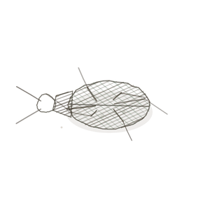
field collected specimen
How to know it on sight
- Simple squamous, one cell thin, with nucleiClusters of neurons in the CNS responsible for processing information. bulging into the lumenThe inside space of a hollow organ or structure.. It looks like a capillary, and it is meant to.
- No brush borderA dense layer of microvilli on the surface of certain cells, increasing surface area for absorption.. Nothing on the surface at all.
- Found only in the medulla, among tubules all cut the same way.
- Tell it from a capillary by what is inside it: a vas rectumThe final section of the large intestine, where feces are stored before elimination. has red cellsThe basic structural and functional units of life., a thin limb is empty.

Specimen 1

Thin limbs among thicker ones. The thin-walled profiles are the descending limbs; the cuboidal ones are already ascending.
Specimen 2

The whole medullary field. Count how many different calibres of tube you can find in one view.
Specimen 3

A vessel, for contrast. Thick wall, a real lumen, and blood in it — none of which a thin limb has.


People keep calling her by the wrong name
Look-alikes
Two plates. Both are thin-walled tubes in the medulla, and only one of them is a tubule.
A vessel in the medulla

Look inside. Red cells mean a vas rectum, and a vas rectum is doing the opposite job — carrying the salt away rather than leaving it behind.
Collecting duct

Wider, paler, and outlined. A collecting duct keeps its sharp borders even when it is squashed flat.
Examine it yourself
The virtual slide
Find the medulla and go as deepAway from the surface of the body. toward the papilla as the section A cut or slice of the body or an organ for study. lets you. Then pick one thin-walled profile and decide, from the contents alone, whether it is a tubule or a capillary.
Virtual slide courtesy of Histology Guide.
YOur missions
Field Notes for Future Missions
Mission 1: label the loop
Mission 2: quick check
Field notes
Why this organism
A heron stands in cold water for hours and does not lose its heat, because the artery going down its leg runs right beside the vein coming back up, and the heat crosses over instead of leaving. Same arrangement, same arithmetic, different cargo: the loop swaps salt and water where the heron swaps heat. It is worth saying out loud that countercurrent exchange was not invented by the kidney — it is a trick that turns up wherever two flows run antiparallel, and a bird the students have watched in a marsh is the cheapest way to make that land.
Next · Pl. V
Tubulus densus var. mimosae
the sensor · distal tubule and macula densaA group of specialized cells in the distal convoluted tubule that detect sodium concentration and he.
In the wild · photograph to come.
Hop to:
← Epithelial Tissues: A Guided Journey
All plates — The Tissue Field Guide: Epithelium
Frontispiece Ordo Epithelia (main page)
Pl. I · Simple squamous
Pl. II · Simple cuboidal
Pl. III · Simple columnar
Pl. IV · Stratified squamous
Pl. V · Stratified cuboidal
Pl. VI · Stratified columnar
Pl. VII · Transitional
Pl. VIII · Pseudostratified columnar
Epithelial Identification Game
All Modules
- Anatomical Language, Membranes & Homeostasis
- Just Enough Chemistry
- The Cell & Its Transport
- Making Cells & Proteins
- The Integumentary System
- The Skeletal System
- The Muscular System
- Nervous Tissue & the Senses
- The Spinal Cord
- The Brain & the Blood–Brain Barrier
- The Autonomic Nervous System
- The Special Senses
- The Endocrine System
- Blood
- The Heart
- Blood Vessels
- The Digestive System
- The Respiratory System
- The Urinary System
- Fluids, Electrolytes & Acid–Base Balance
- The Reproductive System
- The Immune System
List of terms
- nephron
- juxtamedullary
- water
- urine
- collecting duct
- nuclei
- lumen
- brush border
- rectum
- cells
- deep
- section
- macula densa