Time To Read
Date Last Modified
Pl. VI · FAM. GIGNENTIDAE
Gignens medullaris var. termitis
the nursery in the wall · red bone marrow
The plate
The specimen, drawn
Pl. VI

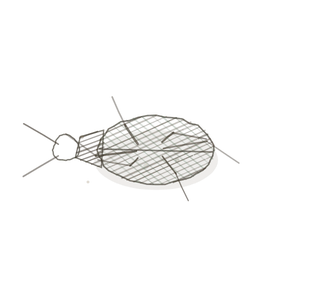
The termite mound as it was drawn, cut open, with red bone marrow below it and a megakaryocyte in the circle.

Habitat & range
Where it lives
Body map

Inside the bones: in an adult, mostly the bones of the trunk.
Range
- In the spaces of spongy bone: sternum, ribs, vertebrae, pelvis, skull.
- In the upper ends of the femur and the humerus.
- In a newborn, in every bone. With age the marrow of the limb bones turns to fat, and is then called yellow marrow.
- Sampled from the back of the hip bone, which is thick, close to the skin and nowhere near anything vital.
Collection record
- Add your slide numbers here.
Niche
What it does for a living
Make every cell in the blood, and make the body’s B lymphocytes ready for work. Red marrow belongs in this volume because it is a primary lymphoid organ: all lymphocytes begin here, B cells finish their training here, and the cells that will become T cells leave from here for the thymus (Pl. V). It is also where red cells, platelets and every other white cell are made, a few hundred billion cells a day, all descended from one kind of stem cell. On a slide it has three parts. The blood-forming cells are packed in cords: cells of every lineage at every stage of maturity, all mixed together, which is why the field looks so busy and so varied. Between the cords run sinusoids, wide capillaries with very thin, leaky walls, and a finished cell leaves by squeezing through that wall into the blood. Scattered among everything are fat cells, more of them with every decade. One cell stands out from the rest: the megakaryocyte, many times the size of its neighbours, with a single large lobed nucleus. It lies against a sinusoid and reaches long processes through the wall, and the blood flowing past breaks pieces off them. Those pieces are the platelets.
field collected specimen
How to know it on sight
- Inside bone. Look for a bony trabecula or the wall of the marrow cavity at the edge of the field.
- Fat cells: large, round, empty-looking rings, scattered through the tissue.
- Between the fat, a crowd of cells that are all different: big and small, dark and pale nuclei, round, lobed and bean-shaped, with red cells among them.
- Megakaryocytes: a few giants, each with one big, many-lobed nucleus. Nothing else on the slide is that size.

Specimen 1

A sinusoid. A wide, thin-walled channel among the blood-forming cells: this is the door the finished cells leave by.
Specimen 2

A megakaryocyte and the platelets that come off it, drawn.
Specimen 3

One of the products, out in the blood: a small lymphocyte among red cells, in a smear.


People keep calling her by the wrong name
Look-alikes
Two other tissues that look like this at a glance: one for the fat, one for the red.
Adipose tissue

Adipose tissue — the same empty rings, but with only thin strands of connective tissue between them. No crowd of blood-forming cells, and no bone round it.
Spleen

Spleen — a red, cellular field full of blood, but with round islands of white pulp in it and no fat cells or megakaryocytes. See Pl. IV.
Examine it yourself
The virtual slide
At low power, find the bone and then the marrow inside it. Estimate how much of the field is fat and how much is cells. Go up on the cells and notice that no two neighbours look the same. Then hunt for the largest cell in the field, and check its nucleus: one nucleus, with many lobes.
Virtual slide courtesy of Histology Guide.
YOur missions
Field Notes for Future Missions
Mission 1: label the marrow
Mission 2: quick check
Field notes
Why this organism
A termite mound is as hard as a brick. You could stand on it. Everything that matters is inside: a maze of chambers and galleries, and in one room near the base, a single enormous queen. She does not leave that room. She lays an egg every few seconds for years, and every other animal in the mound, a million or more, came out of her. They do not all turn out alike. Most become workers, small and plain. Some become soldiers, with heavy heads and jaws. A few grow wings, and on one warm evening they pour out of the top of the mound and are gone. Three body plans for three jobs, from one source, made inside a hard wall and released when they are finished. That is red marrow. It lies inside bone, the hardest tissue you have. It turns out several entirely different kinds of cell: red cells to carry oxygen, white cells to fight, platelets to plug leaks. All of them descend from one kind of stem cell, and none of them is made anywhere else. The finished cells leave through the wall of a sinusoid, and most never come back. Two honest differences. The mound has one queen, and marrow has many thousands of stem cells, spread through the skeleton, so no single loss ends the supply. And a termite’s caste is settled by how it is fed and by the scents round it as it grows, while a blood cell’s fate is settled by chemical signals, hormones such as erythropoietin, that rise and fall with what the body is short of. Lose blood, and within days the marrow is making more red cells. The mound cannot do that so quickly.
Next · Pl. VII
Vigilans appendicis var. abelmoschi
the dead end · appendix.
In the wild · photograph to come.
Hop to:
← Epithelial Tissues: A Guided Journey
All plates — The Tissue Field Guide: Epithelium
Frontispiece Ordo Epithelia (main page)
Pl. I · Simple squamous
Pl. II · Simple cuboidal
Pl. III · Simple columnar
Pl. IV · Stratified squamous
Pl. V · Stratified cuboidal
Pl. VI · Stratified columnar
Pl. VII · Transitional
Pl. VIII · Pseudostratified columnar
Epithelial Identification Game
All Modules
- Anatomical Language, Membranes & Homeostasis
- Just Enough Chemistry
- The Cell & Its Transport
- Making Cells & Proteins
- The Integumentary System
- The Skeletal System
- The Muscular System
- Nervous Tissue & the Senses
- The Spinal Cord
- The Brain & the Blood–Brain Barrier
- The Autonomic Nervous System
- The Special Senses
- The Endocrine System
- Blood
- The Heart
- Blood Vessels
- The Digestive System
- The Respiratory System
- The Urinary System
- Fluids, Electrolytes & Acid–Base Balance
- The Reproductive System
- The Immune System