Time To Read
Date Last Modified
Pl. I · FAM. PERIPHERICIDAE
Periphericus fasciculatus var. porraceus
the cable bundle · peripheral nerve
The plate
The specimen, drawn
Pl. I

The tissue as you will meet it on the slide.

Habitat & range
Where it lives
Body map

Where this tissue is found.
Range
- Every nerve outside the brain and the cord — which is all of them.
- The brachial plexusA network of nerves in the neck and shoulder that control the upper limbs., and the nerves that run from it down the arm.
- The sciatic nerveThe largest nerve in the body, arising from the sacral plexus., the thickest in the body, hip to foot.
- Twelve pairs of cranial nervesNerves that arise from the brain and control head and neck functions., leaving the skull through their own holes.
- And the roots, where they leave the cord — one pair in, one pair out, every segment.
Collection record
- Add your slide numbers here.
Niche
What it does for a living
Carrying, in both directions, over distance. A peripheral nerve is not one cell and not one signal: it is a bundle of axons travelling together, some bringing sensation in and some taking orders out, wrapped in three nested sheaths that keep them insulated, fed and mechanically safe. Myelin supplies the speed; the sheaths supply the engineering. Cut one and the axons can regrow along the tubes those sheaths leave behind, which is why a peripheral nerve can recover and a tract inside the cord cannot.
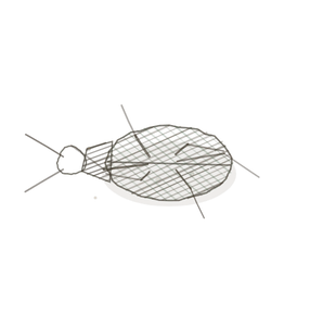
field collected specimen
How to know it on sight
- Cut across, circles within circles — each axon a dark dot inside a pale ring of myelin.
- The axons are gathered into fasciclesBundles of nerve fibers within a nerve.
or
Bundles of nerve fibers within a muscle., and the fascicles gathered into a trunk. - Three sheaths, nested: endoneuriumA thin layer of connective tissue surrounding each nerve fiber. round each axon, perineuriumA connective tissue sheath that surrounds a bundle (fascicle) of nerve fibers. round each fascicle, epineuriumConnective tissue sheath surrounding an entire nerve. round the whole nerve.
- Cut lengthwise the fibres run wavy and parallel, with the occasional pinch of a node.

Specimen 1

The three sheaths, outside in. Find the outermost wrapper first, then work your way down.
Specimen 2

Both cuts in one field — round profiles where the knife went across, wavy lines where it went along.
Specimen 3

Cut across at high power. Every pale ring is myelin, and the dark dot inside it is the axon.


People keep calling her by the wrong name
Look-alikes
Two plates. Read the line underneath each one before you decide.
Smooth muscle

Tell them apart by the rings. Smooth muscle is solid pink with nucleiClusters of neurons in the CNS responsible for processing information. in it; nerve is full of pale rings with a dot in the middle of each.
Dense regular connective tissue

Tell them apart lengthwise. Tendon is straight parallel bundles; nerve fibres are wavy, and they travel in fascicles with a sheath around each.
Examine it yourself
The virtual slide
Find the edge of one fascicle and trace all the way round it. Then go up in power and look for the pale ring around a single dark axon.
Virtual slide courtesy of Histology Guide.
YOur missions
Field Notes for Future Missions
Mission 1: peel the sheaths
Mission 2: quick check
Field notes
Why this organism
A leek is a stack of leaves pretending to be a stem. Peel it and the layers come away in order, each one having wrapped the one inside it, all the way down to the pale core. A peripheral nerve is built the same way and it is the only reason the vocabulary is learnable: endoneurium round one axon, perineurium round a bundle of them, epineurium round the whole cable. Three words, one prefix each, and the prefixes are doing all the work. Cut a leek across and the rings tell you the same story the slide does.
Next · Pl. II
Periphericus globosus var. allianus
the garlic bulb · a ganglionA cluster of neuron cell bodies located in the peripheral nervous system (PNS)..
Hop to:
← Epithelial Tissues: A Guided Journey
All plates — The Tissue Field Guide: Epithelium
Frontispiece Ordo Epithelia (main page)
Pl. I · Simple squamous
Pl. II · Simple cuboidal
Pl. III · Simple columnar
Pl. IV · Stratified squamous
Pl. V · Stratified cuboidal
Pl. VI · Stratified columnar
Pl. VII · Transitional
Pl. VIII · Pseudostratified columnar
Epithelial Identification Game
All Modules
- Anatomical Language, Membranes & Homeostasis
- Just Enough Chemistry
- The Cell & Its Transport
- Making Cells & Proteins
- The Integumentary System
- The Skeletal System
- The Muscular System
- Nervous Tissue & the Senses
- The Spinal Cord
- The Brain & the Blood–Brain Barrier
- The Autonomic Nervous System
- The Special Senses
- The Endocrine System
- Blood
- The Heart
- Blood Vessels
- The Digestive System
- The Respiratory System
- The Urinary System
- Fluids, Electrolytes & Acid–Base Balance
- The Reproductive System
- The Immune System
List of terms
- brachial plexus
- sciatic nerve
- cranial nerves
- fascicles
- endoneurium
- perineurium
- epineurium
- nuclei
- ganglion