Time To Read
Date Last Modified
Pl. IV · FAM. CENTRALIDAE
Centralis corticalis var. lichenosus
the crust · cerebrum
The plate
The specimen, drawn
Pl. IV

The tissue as you will meet it on the slide.

Habitat & range
Where it lives
Body map

Where this tissue is found.
Range
- The entire folded surface of both hemispheres — a sheet only a few millimetres thick.
- Folded into gyri and sulci, so that most of it is hidden down inside the folds.
- Plus deepAway from the surface of the body. islands of grey, the basal nucleiClusters of neurons in the brain that help regulate movement and coordination., marooned inside the white matterThe outer portion of the spinal cord made of myelinated nerve fibers that transmit signals..
Collection record
- Add your slide numbers here.
Niche
What it does for a living
Everything that is not automatic. The cerebral cortexThe outermost layer of the brain, responsible for higher-order thinking and processing. is where sensation becomes perception, where a movementA fundamental property of life involving motion of the body or its parts. is decided rather than executed, and where language, memoryThe ability to store and recall information. and judgement live. It is built as a sheet because a sheet lets neuronsThe functional cells of the nervous system that transmit signals. stand side by side in columns and talk to their neighbours; it is folded because the sheet is far too big for the skull it has to fit inside. Spread flat it would cover a small table.
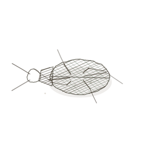
field collected specimen
How to know it on sight
- A darker band of cell bodies following the surface, with pale fibre underneath it.
- That band is only a few millimetres thick, and its deep edge is surprisingly abrupt.
- Scattered through it, triangular pyramidal cellsLarge neurons in the cerebral cortex involved in voluntary motor control. with their points aimed at the surface.
- Six layers if you look hard — but they shade into one another, nothing like the cerebellum’s three.

Specimen 1

Two gyri at low power. The darker band follows the folded surface; the pale core is white matter.
Specimen 2

A whole slab. Grey on the outside, white within — hold on to that and Pl. V will catch you out less.
Specimen 3

One gyrus close up, with a sulcus beside it. The cortex dives down into the fold and keeps going.


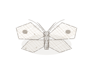
People keep calling her by the wrong name
Look-alikes
Two plates. Read the line underneath each one before you decide.
Cerebellum

Tell them apart by the row. The cerebellumThe brain region responsible for balance, coordination, and fine motor control. has three sharp layers and a single row of giant cellsThe basic structural and functional units of life.; the cerebrum has neither.
Grey matter of the spinal cord

Tell them apart by where the grey is. Here it is a band on the outside; in the cord it is a butterfly in the middle.
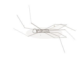
Examine it yourself
The virtual slide
Find the outer surface, then follow the dark bandThe part of the sarcomere containing thick filaments. inward until it stops. That stopping point is the boundary — and it is the whole exercise.
Virtual slide courtesy of Histology Guide.
YOur missions
Field Notes for Future Missions
Mission 1: find the edge
Mission 2: quick check
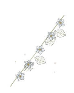
Field notes
Why this organism
A foliose lichen is a crust with a cortex — and that is not a metaphor borrowed for the occasion. Cortex is the actual term for the dense protective layer on the upper surface of a lichen thallus. Cut one across and you find a tough cortex on top, a bright layer of algal cells just beneath it where all the work happens, and a pale medulla of loose fungal thread filling the middle. A slice of cerebrum reads exactly the same way: cell bodies crowded at the surface, the working layer, and pale myelinated thread underneath. Anatomy borrowed the word, and it borrowed it from something very like this.
Next · Pl. V
Centralis inversus var. interrogationis
the butterfly · the spinal cordThe central nervous system structure that relays signals between the brain and body..
Hop to:
← Epithelial Tissues: A Guided Journey
All plates — The Tissue Field Guide: Epithelium
Frontispiece Ordo Epithelia (main page)
Pl. I · Simple squamous
Pl. II · Simple cuboidal
Pl. III · Simple columnar
Pl. IV · Stratified squamous
Pl. V · Stratified cuboidal
Pl. VI · Stratified columnar
Pl. VII · Transitional
Pl. VIII · Pseudostratified columnar
Epithelial Identification Game
All Modules
- Anatomical Language, Membranes & Homeostasis
- Just Enough Chemistry
- The Cell & Its Transport
- Making Cells & Proteins
- The Integumentary System
- The Skeletal System
- The Muscular System
- Nervous Tissue & the Senses
- The Spinal Cord
- The Brain & the Blood–Brain Barrier
- The Autonomic Nervous System
- The Special Senses
- The Endocrine System
- Blood
- The Heart
- Blood Vessels
- The Digestive System
- The Respiratory System
- The Urinary System
- Fluids, Electrolytes & Acid–Base Balance
- The Reproductive System
- The Immune System
List of terms
- cerebrum
- deep
- basal nuclei
- white matter
- cerebral cortex
- movement
- memory
- neurons
- pyramidal cells
- cerebellum
- cells
- dark band
- spinal cord