Time To Read
Date Last Modified
Pl. III · FAM. NEPHRONIDAE
Tubulus villosus var. callistemonis
the greedy one · proximal convoluted tubuleThe first part of the nephron tubule where most reabsorption of water, ions, and nutrients occurs.
The plate
The specimen, drawn
Pl. III

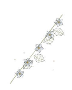
The flower as it was drawn, with the tubule below it and the brush borderA dense layer of microvilli on the surface of certain cells, increasing surface area for absorption. in the circle.

Habitat & range
Where it lives
Body map

Cortex, out in the labyrinth, coiled tight around its own corpuscle.
Range
- Cortex, in the labyrinth, coiled tight around the corpuscle it came from.
- The most numerous profile in a cortical field, by a wide margin.
- Takes back 65 % of the filtrateThe fluid that is filtered from the blood into the nephron and will eventually become urine. before anything else gets a turn.
- Where all of the glucoseA simple sugar that is the main source of energy for cells. and all of the amino acids go, and where most drugs are secreted.
Collection record
- Add your slide numbers here.
Niche
What it does for a living
Bulk recovery, and it is not subtle about it. Two thirds of the waterThe universal solvent essential for life., two thirds of the sodium(Na⁺): Major ECF cation; important for fluid balance, nerve function., all of the glucose, all of the amino acids, most of the bicarbonate(HCO₃⁻) – A crucial buffer in blood that helps maintain pH balance; formed when carbon dioxide react — reclaimed here, before any hormone has a say. It is not regulated minute to minute; it simply takes, which is why the transporters saturate and why glucose appears in the urineThe liquid waste excreted by the kidneys. above a plasmaThe liquid component of blood. level of about 180 mg/dL. The brush border is the reason it can: microvilliTiny projections on the surface of epithelial cells that increase surface area for absorption. multiply the surface about twenty-fold, and the mitochondria behind them pay for the pumping. It is also the first thing to die when the kidney loses its blood supply, because it is the most expensive tissue in the organ to run.
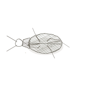
field collected specimen
How to know it on sight
- The lumenThe inside space of a hollow organ or structure. is never clean. A brush border of microvilli fills it with fuzz. This is the diagnosis, and it beats everything else in the kidney.
- Cells tall, cytoplasmThe gel-like substance within a cell that contains organelles and cytosol. deeply eosinophilic — the pinkest thing in the field, because they are packed with mitochondria.
- Cell borders indistinct. You often cannot tell where one cell stops and the next starts.
- Round nucleiClusters of neurons in the CNS responsible for processing information., widely spaced, and fewer of them around a profile than the tubule next door.

Specimen 1

Pick one profile and try to draw the line between two of its cellsThe basic structural and functional units of life.. The difficulty is the finding.
Specimen 2

Compare the cytoplasm here with the paler tubules around it. Pink means mitochondria, and mitochondria mean pumping.
Specimen 3

Cut obliquely, the brush border smears across the lumen instead of ringing it. Same tissue, different angle.


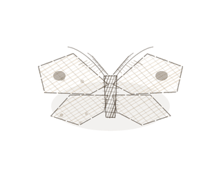
People keep calling her by the wrong name
Look-alikes
Two plates. Both are tubules in the same field, and neither one has a brush border.
Distal tubule

A clean, open lumen and more nuclei around the ring. If you can see into it clearly, it is not proximalCloser to the point of attachment or origin..
Collecting duct

Pale cytoplasm and borders you could cut yourself on. Never sort these three by diameter — sort them by the lumen and the borders.
Examine it yourself
The virtual slide
Go to a cortical view at the highest magnification and find two tubule profiles touching each other, one with a lumen you can see into and one without. Then say out loud which is which, and why.
Virtual slide courtesy of Histology Guide.
YOur missions
Field Notes for Future Missions
Mission 1: label the wall
Mission 2: quick check
Field notes
Why this organism
A bottlebrush is a dense fuzz of filaments filling all the space around a central stalk, so you never see the stalk cleanly — which is precisely what stops you seeing a proximal tubule’s lumen. The flower makes the functional point too: all that fuzz is surface area, deployed for the sole purpose of catching things that pass by. Nothing about a bottlebrush is restrained, and nothing about a proximal tubule is either.
Next · Pl. IV
Tubulus tenuis var. ardeae
the long game · nephronThe functional unit of the kidney that filters blood and produces urine. loop, thin limb.
In the wild · photograph to come.
Hop to:
← Epithelial Tissues: A Guided Journey
All plates — The Tissue Field Guide: Epithelium
Frontispiece Ordo Epithelia (main page)
Pl. I · Simple squamous
Pl. II · Simple cuboidal
Pl. III · Simple columnar
Pl. IV · Stratified squamous
Pl. V · Stratified cuboidal
Pl. VI · Stratified columnar
Pl. VII · Transitional
Pl. VIII · Pseudostratified columnar
Epithelial Identification Game
All Modules
- Anatomical Language, Membranes & Homeostasis
- Just Enough Chemistry
- The Cell & Its Transport
- Making Cells & Proteins
- The Integumentary System
- The Skeletal System
- The Muscular System
- Nervous Tissue & the Senses
- The Spinal Cord
- The Brain & the Blood–Brain Barrier
- The Autonomic Nervous System
- The Special Senses
- The Endocrine System
- Blood
- The Heart
- Blood Vessels
- The Digestive System
- The Respiratory System
- The Urinary System
- Fluids, Electrolytes & Acid–Base Balance
- The Reproductive System
- The Immune System
List of terms
- proximal convoluted tubule
- brush border
- filtrate
- glucose
- water
- sodium
- bicarbonate
- urine
- plasma
- microvilli
- lumen
- cytoplasm
- nuclei
- cells
- proximal
- nephron