Time To Read
Date Last Modified
Frontispiece · ORD. GENERATIVA
Ordo Generativa
the makers · the reproductive systemThe organ system responsible for producing gametes and offspring.
The plates
The eleven specimens
Five families, and the split between them is the lesson. Three plates make gametes, one ripens and stores them, two carry them, three make the fluid they travel in, and two receive — and only the first three have germ cellsThe basic structural and functional units of life. anywhere in the wall.
Pl. I
Generans gradatus var. helianthus
The Ripening Wheel · testis

Open Pl. I →
Pl. II
Maturans stereociliatus var. gordius
The Long Wait · epididymis

Open Pl. II →
Pl. III
Vehens muscularis var. caryae
The Pinhole Cannon · ductus deferens

Open Pl. III →
Pl. IV
Glandula concretionis var. crassostrea
The Pearl-Keeper · prostate

Open Pl. IV →
Pl. V
Glandula labyrinthica var. clathratus
The False Rooms · seminal vesicle

Open Pl. V →
Pl. VI
Glandula lubricans var. myxinus
The Slipstream · bulbourethral gland

Open Pl. VI →
Pl. VII
Generans follicularis var. nymphaeus
The All-At-Once · ovary

Open Pl. VII →
Pl. VIII
Generans luteus var. nymphaeus senescens
The Afterglow · corpus luteum

Open Pl. VIII →
Pl. IX
Vehens ciliatus var. crinoideus
The Feather Conveyor · uterine tube

Open Pl. IX →
Pl. X
Recipiens cyclicus var. acerinus
The Sap Run · uterusThe muscular organ where a fertilized egg implants and develops.

Open Pl. X →
Pl. XI
Recipiens transiens var. balanus
The Borderline · cervix

Open Pl. XI →
The ground plan
Lining plus wall
Every organ in this volume is a tube, and you read each one from two things: what the lining is doing, and how much muscle is wrapped around it. The lining is the first question because it is the one that cannot be faked by a bad section A cut or slice of the body or an organ for study.. The wall is the second because two organs here share a lining and differ only in their wall. In the female tract there is a third question, and it is not anatomical at all — it is which week of the cycle you are looking at.
Fig. 1 · lining plus wall

One tube in section, the five linings of this volume ranged around it, and the wall thickness called out on its own — because wall is what separates the two organs the lining cannot.
The five words
- Lumen — the space inside. Its shape and what is floating in it both count as evidence.
- Lining — the epithelium facing the lumenThe inside space of a hollow organ or structure.. Five of them in this volume, and naming the lining gets you most of the way to naming the organ.
- Muscularis — the muscle coat around the lining. Thin, thick, or three layers thick, and it is measured against the lumen, never on its own.
- Stroma — the connective tissue the whole thing sits in. In the ovary and the endometriumThe inner lining of the uterus that thickens during the menstrual cycle to support a potential pregn it is not packing material, it is the thing you are reading.
- Gamete — an egg or a sperm. Where they are tells you the family: in the wall means a gonad, in the lumen means a duct carrying cargo.
Name the lining, then measure the wall against the lumen. Those two in that order settle most of this volume.

First question
Are there germ cells in the wall?
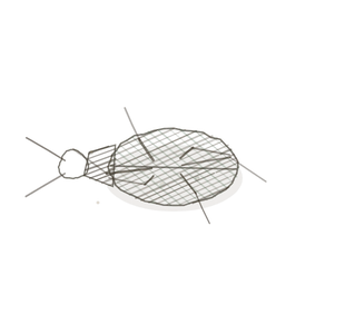
Before anything else, decide whether the wall itself is making cells or merely moving them. A gonad has germ cells at several stages built into its wall. A duct has one tidy epithelium and whatever it happens to be carrying.
Fig. 2 · a gonad wall, and a duct wall

Two walls side by side. On the left, several cell generations stacked from the outside in. On the right, a single row of cells and the cargo sitting loose in the lumen.
- A gonad — the wall is several cells deepAway from the surface of the body. and the cells are visibly different from each other, because they are at different stages. Testis and ovary both qualify, in their own ways.
- A duct or a gland — one epithelium, all of its cells alike, and anything else in the field is either cargo in the lumen or secretionThe process of moving substances from the blood into the nephron tubule to be excreted in urine..
- The trap — an epididymal lumen packed solid with sperm. That is a duct with a full load, not a gonad. The sperm are cargo, not residents.
Germ cells in the wall means a gonad. Sperm in the lumen means nothing at all about the family — ducts are full of them by design.
Second question
What is the lining?
There are five linings in this volume and they are genuinely distinguishable at 40×. Learn them as a row and most of the plates name themselves.
Fig. 3 · the five linings

Five linings drawn flat in a row, in the order you are most likely to meet them.
- Germinal stratified — several generations of cells from the outer wall inward, getting smaller and darker as they go. Only the testis looks like this.
- Pseudostratified columnar with stereocilia — tall cells with long still tufts on top, and nucleiClusters of neurons in the CNS responsible for processing information. at two heights that make it look layered when it is not. Epididymis and ductus deferens both. The wall decides which.
- Ciliated simple columnar with peg cells — one row of tall cells with true ciliaHair-like projections on the surface of some cells that move fluids or particles., interrupted by short wedge-shaped cells with no cilia at all. The uterine tube, and nothing else here.
- Secretory simple columnar — one row of plain tall cells with secretion in or above them. The glands and the endometrium.
- Stratified squamous — many layers, flattening towards the surface. The cervix at its outer end, where it meets the vaginaThe muscular canal connecting the uterus to the external genitalia..
Stereocilia are not cilia. They are long microvilliTiny projections on the surface of epithelial cells that increase surface area for absorption., they do not move, and nothing in the male duct system is ciliated — so a visibly ciliated border puts you in the uterine tube.

The naming key
How to read a name
Three parts. The genus says what the tube is for; the species says what the lining is doing; the variety is the organismA living individual made up of cells. the plate is drawn as, and carries no anatomyThe study of the structure of the human body. at all.
Genus · what the tube is for
- Generans — gamete-making. Germ cells at stages in the wall itself.
- Maturans — ripening and storing what arrives already made.
- Vehens — carrying.
- Glandula — secreting. No gamete A haploid reproductive cell (sperm or egg). ever passes through.
- Recipiens — receiving.
Species · what the lining is doing
- gradatus — in stages, ranged across the wall
- stereociliatus — tufted, and the tufts do not move
- muscularis — the wall is the whole point
- concretionis — secretion that sets hard
- labyrinthica — folded until one chamber reads as many
- lubricans — making slip
- follicularis — each gamete in its own chamber
- luteus — the chamber after the gamete has gone
- ciliatus — truly ciliated, and sweeping
- cyclicus — the same tissue, different every week
- transiens — one lining becoming another
Worked both ways
- Vehens muscularis → carrying, and the wall is the whole point → a pinhole lumen in an enormous three-layer wall → ductus deferens.
- Generans follicularis → gamete-making, each one in its own chamber → folliclesStructures in the ovaries that contain developing oocytes. at several sizes in a dense stroma → ovary.
- You saw one row of tall ciliated cells with short wedge cells between them, in a wall thrown into deep folds → truly ciliated and sweeping → Vehens ciliatus → the uterine tube.
Three that trip people up
The look-alikes
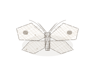
Three pairs account for most of the mistakes in this volume. Each has one reliable test, and in two of the three the lining is identical, so the lining is not it.
Fig. 4 · three that trip people up

Three pairs, stacked. Lining, then wall, then what is in the lumen.
An epididymis, or a ductus deferens?
The trap: the lining is the same in both — pseudostratified columnar with stereocilia.
They sit next to each other in the tract and the epithelium will not separate them, because it is genuinely the same epithelium. Lumen contents will not do it either; both can be full of sperm.
Measure the wall against the lumen. A wide lumen in a thin wall is epididymis. A pinhole lumen in a wall three muscle layers thick is ductus deferens. The ratio is not subtle once you look for it.
A prostate, or a seminal vesicle?
The trap: both are folded glands with a secretory columnar lining, sitting side by side on the slide.
Both throw their mucosaThe innermost lining of the digestive tract that contains mucus-secreting cells for protection and a into folds and both are packed with secretion, so neither folding nor secretion decides it.
Look for stones. Round laminated concretions in the lumen mean prostate. Honeycombed folds that fuse into a lattice of false chambers, with no stones, mean seminal vesicle.
Proliferative endometrium, or secretory?
The trap: it is the same organ, the same lining and the same stroma, twice.
Nothing anatomical separates these, because there is nothing anatomical to separate. The difference is which hormone was dominant that week.
Read the glands. Straight and narrow with a thin stroma means proliferative. Coiled and saw-toothed, with a thick oedematous stroma and prominent spiral arteriesBlood vessels supplying the endometrium, shedding during menstruation., means secretory.
Keying out a specimen
Four questions, in this order
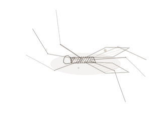
This is the procedure. It works on a reproductive slide you have never seen before and have not been told the name of, which is the only test that matters.
- Are there germ cells at different stages in the wall? — Yes, and in round tubule profiles, means testis. Yes, in whole chambers of different sizes, means ovary. No means a duct or a gland, and you carry on.
- What is the lining? — Pick one of the five. If it is ciliated with peg cells you are finished: uterine tube. If it is stratified squamous you are at the cervix or beyond.
- How much muscle, relative to the lumen? — A pinhole lumen in an enormous three-layer wall is the ductus deferens and almost nothing else. This is the question that separates the pair the lining cannot.
- If it is female — what phase is it in? — Straight narrow glands mean proliferative. Coiled saw-toothed glands with prominent spiral arteriesBlood vessels that carry oxygenated blood away from the heart (except pulmonary arteries, which carr mean secretory. The question only applies to the endometrium, and on the endometrium it is the whole answer.
Three overrides, and students get all three wrong. Germ cells in the wall mean a gonad, not a duct — an epididymal lumen packed with sperm is still a duct, because the sperm are cargo. Stereocilia are not cilia — they do not move, and nothing in the male duct system is ciliated, so visible cilia put you in the uterine tube. A star-shaped lumen in a thick muscular wall is not automatically a ureterThe tube that carries urine from the kidney to the bladder. — check the lining, because urothelium means ureter and pseudostratified columnar means ductus deferens.
YOur missions
Field Notes for Future Missions
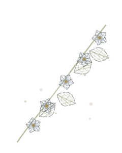
Before you go to the plates, try the key on the whole tract at once.
Mission 1 · follow a gamete out
Mission 2 · key it out
Field notes
Why eleven organisms
A sunflower headRounded proximal end that fits into the acetabulum of the hip bone., a horsehair worm, a shagbark hickory nut, a feather star, an eastern oyster, a lattice stinkhorn, a hagfish, a fragrant waterThe universal solvent essential for life. lily, the same lily spent, a sugar maple and an acorn barnacle have nothing to do with one another and nothing to do with reproductive tractsBundles of nerve fibers in the CNS that carry signals between brain regions.. That is rather the point of them.
A list of eleven structures is something you memorise and then lose. Eleven organisms that each carry one structural idea — stages ranged in a wheel from rim to centre, an enormous length folded into a small space, a pinhole cavity inside a massive husk, ciliated arms sweeping cargo along, a secretion that sets into a stone, a lattice of chambers that are mostly empty space, slip made on demand, one chamber opening at a time, the chamber left behind after it closes, the same tissue running or still depending on the season, and a hard plate meeting soft tissue at an abrupt line — stay put, because you can picture them. The name is a handle for the picture, not the other way round.
Begin · Pl. I
Generans gradatus
The Ripening Wheel · testis.
Hop to:
← Epithelial Tissues: A Guided Journey
All plates — The Tissue Field Guide: Epithelium
Frontispiece Ordo Epithelia (main page)
Pl. I · Simple squamous
Pl. II · Simple cuboidal
Pl. III · Simple columnar
Pl. IV · Stratified squamous
Pl. V · Stratified cuboidal
Pl. VI · Stratified columnar
Pl. VII · Transitional
Pl. VIII · Pseudostratified columnar
Epithelial Identification Game
All Modules
- Anatomical Language, Membranes & Homeostasis
- Just Enough Chemistry
- The Cell & Its Transport
- Making Cells & Proteins
- The Integumentary System
- The Skeletal System
- The Muscular System
- Nervous Tissue & the Senses
- The Spinal Cord
- The Brain & the Blood–Brain Barrier
- The Autonomic Nervous System
- The Special Senses
- The Endocrine System
- Blood
- The Heart
- Blood Vessels
- The Digestive System
- The Respiratory System
- The Urinary System
- Fluids, Electrolytes & Acid–Base Balance
- The Reproductive System
- The Immune System
List of terms
- reproductive system
- cells
- uterus
- section
- lumen
- endometrium
- deep
- secretion
- nuclei
- cilia
- vagina
- microvilli
- organism
- anatomy
- gamete
- follicles
- mucosa
- spiral arteries
- arteries
- ureter
- head
- water
- tracts