Time To Read
Date Last Modified
Pl. XI · FAM. RECIPIENTIDAE
Recipiens transiens
The Borderline · cervix
The plate
The specimen, drawn
Pl. XI

The rock at low tide, with the transformation zone below it and the junction read at ×200 in the circle.

Habitat & range
Where it lives

Range
- The lower third of the uterus, projecting into the vault of the vagina.
- About 2.5 cm long, with a narrow canal running through it between the internal and external os.
- The canal is lined by mucus-secreting simple columnar; the part facing the vagina by stratified squamous.
- Where those two linings meet is the transformation zone, and its position shifts with age and hormonal state.
Collection record
- Add your slide numbers here.
Niche
What it does for a living
Be a door. Mucus-plugged and hostile for most of the month, thin and passable for a few days around ovulationThe release of a mature oocyte from the ovary., and tough enough below to survive being a passage.
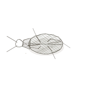
field collected specimen
How to know it on sight
- Two completely different linings meeting at a line, with no gradient between them.
- On the canal side, tall pale mucus-secreting simple columnar, often with branching crypts dipping into the stroma.
- On the vaginal side, non-keratinised stratified squamous many cellsThe basic structural and functional units of life. deepAway from the surface of the body., nucleiClusters of neurons in the CNS responsible for processing information. flattening toward the surface.
- The basement membrane runs straight on underneath both. The epithelium changes; the membrane does not.

Specimen 1

The cervix at higher power — the lining and the stroma beneath it, with blood and surface cells.
Specimen 2

Cervical stratified squamous at high power. Well-ordered layers above, flattening as they approach the surface.
Specimen 3

Transitional epithelium, for contrast: another multi-layered lining that is not squamous and belongs elsewhere.


People keep calling her by the wrong name
Look-alikes
Two plates. The first is the lining below the line on its own, which is what you will see if your section A cut or slice of the body or an organ for study. misses the junction. The second is the organ above, which shares the columnar lining but cycles and sheds, and this one does neither.
Vagina / ectocervix
Many layers, flattening toward the surface, no glands and no columnar anywhere. A section this uniform has missed the zone.
Uterus

Simple columnar again, but making tubular glands in a layer that will be shed. The cervix has crypts and sheds nothing.
Examine it yourself
The virtual slide
Zoom to ×40 and run along the surface until the epithelium changes. Stop on the change itself: that single line, with the basement membrane running on underneath it, is the whole plate.
Virtual slide courtesy of Histology Guide.
YOur missions
Field Notes for Future Missions
Mission 1: find the line
Mission 2: quick check
Field notes
Why this organism
Walk a rocky shore at low tide and the barnacles stop in a line you could draw with a ruler. Above it they desiccate; below it they are eaten. The line is not a wall, it is where two different sets of rules meet, and it is the most biologically interesting place on the rock. The cervical transformation zone is that line, drawn inside a person: columnar above, squamous below, and the join is where cells get confused and where every screening test aims.
Back · Frontispiece
Ordo Generativa
the makers · the whole volume.
In the wild · photograph to come.
Hop to:
← Epithelial Tissues: A Guided Journey
All plates — The Tissue Field Guide: Epithelium
Frontispiece Ordo Epithelia (main page)
Pl. I · Simple squamous
Pl. II · Simple cuboidal
Pl. III · Simple columnar
Pl. IV · Stratified squamous
Pl. V · Stratified cuboidal
Pl. VI · Stratified columnar
Pl. VII · Transitional
Pl. VIII · Pseudostratified columnar
Epithelial Identification Game
All Modules
- Anatomical Language, Membranes & Homeostasis
- Just Enough Chemistry
- The Cell & Its Transport
- Making Cells & Proteins
- The Integumentary System
- The Skeletal System
- The Muscular System
- Nervous Tissue & the Senses
- The Spinal Cord
- The Brain & the Blood–Brain Barrier
- The Autonomic Nervous System
- The Special Senses
- The Endocrine System
- Blood
- The Heart
- Blood Vessels
- The Digestive System
- The Respiratory System
- The Urinary System
- Fluids, Electrolytes & Acid–Base Balance
- The Reproductive System
- The Immune System
List of terms
- neck
- uterus
- vagina
- ovulation
- cells
- deep
- nuclei
- section