Time To Read
Date Last Modified
Pl. I · FAM. LAXIDAE
Laxus vagus var. tillandsius
the packing peanut · areolar connective tissue
The plate
The specimen, drawn
Pl. I

Loose, open, and going nowhere in particular. Fibres of all three kinds, cellsThe basic structural and functional units of life. scattered through, and more space than anything else.

Habitat & range
Where it lives
Body map

Areolar tissue is the one that is simply everywhere. If you are not standing in some other tissue, you are standing in this.
Range
- Under the whole skinThe body’s largest organ, providing protection and regulation., as the papillary layerhe upper layer of the dermis that contains blood vessels and sensory receptors. of the dermisThe thick inner layer of the skin that contains blood vessels, nerves, and connective tissue..
- Packing the gaps between organs, and between the muscles.
- Wrapping every blood vessel and every nerve on its way through.
- Forming the mucous and serous membranesThin tissues that line body cavities and secrete fluid. with the epithelium above it.
- Anywhere a tissue needs to be held near another without being tied to it.
Collection record
- Add your slide numbers here.
Niche
What it does for a living
Filling the gap without closing it. Areolar tissue holds things near each other while leaving room for everything that has to pass between them — vessels, nerves, wandering immune cells, waterThe universal solvent essential for life.. Most of its volume is not fibre at all but ground substance, and that open space is the function, not a shortfall. It is also where infection gets fought: mast cells, macrophages and plasma cellsImmune cells that develop from B cells and produce antibodies. all live here, waiting.
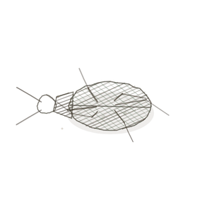
field collected specimen
How to know it on sight
- All three fibre types in one field — pink collagenA structural protein in the dermis that provides strength and elasticity., fine dark elastic, and reticular if the stain shows it.
- No grain. Fibres run every direction, and no direction dominates.
- Cells few and scattered, with wide empty-looking space between everything.
- Thin and pale compared with any dense tissue; you can see through the gaps.

Specimen 1

Areolar tissue in thin skinSkin found on most of the body, lacking a stratum lucidum.. Pick one collagen bundle and follow it until it crosses another going the other way.
Specimen 2

From the scalp. Count how much of this field is fibre and how much is open space — the space wins.

People keep calling her by the wrong name
Look-alikes
Two plates. Read the line underneath each one before you decide.
Dense irregular connective tissue

Both have fibres running every way. Tell them apart by how much space is left: areolar is mostly gap, dense irregular is mostly bundle.
Reticular connective tissue

Reticular is a fine branching net of one fibre type, usually silver-stained dark; areolar is a loose mixture of all three in pink.
Examine it yourself
The virtual slide
Mesentery is areolar tissue spread out thin enough to see through. Find a spot where two collagen bundles cross and look at what is between them — that space is the tissue doing its job.
Virtual slide courtesy of Histology Guide.
YOur missions
Field Notes for Future Missions
Mission 1: all three fibres
Mission 2: quick check
Field notes
Why this organism
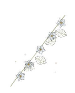
Spanish moss is not a parasite and not a vine. It hangs in the gap between branches, touching everything and attached to almost nothing, and it is mostly air. You cannot pull it tight and you would not want to: the point of it is the space it holds open. Areolar tissue is the same proposition. It fills the room between things without fastening them together, and what passes through that room — blood, lymph, nerves, white cells on patrol — matters more than the fibres themselves.
Next · Pl. II
Laxus adiposus var. mellifer
the vault · adipose tissue.
Hop to:
← Epithelial Tissues: A Guided Journey
All plates — The Tissue Field Guide: Epithelium
Frontispiece Ordo Epithelia (main page)
Pl. I · Simple squamous
Pl. II · Simple cuboidal
Pl. III · Simple columnar
Pl. IV · Stratified squamous
Pl. V · Stratified cuboidal
Pl. VI · Stratified columnar
Pl. VII · Transitional
Pl. VIII · Pseudostratified columnar
Epithelial Identification Game
All Modules
- Anatomical Language, Membranes & Homeostasis
- Just Enough Chemistry
- The Cell & Its Transport
- Making Cells & Proteins
- The Integumentary System
- The Skeletal System
- The Muscular System
- Nervous Tissue & the Senses
- The Spinal Cord
- The Brain & the Blood–Brain Barrier
- The Autonomic Nervous System
- The Special Senses
- The Endocrine System
- Blood
- The Heart
- Blood Vessels
- The Digestive System
- The Respiratory System
- The Urinary System
- Fluids, Electrolytes & Acid–Base Balance
- The Reproductive System
- The Immune System
List of terms
- cells
- skin
- papillary layer
- dermis
- serous membranes
- water
- plasma cells
- collagen
- thin skin