Time To Read
Date Last Modified
Pl. I · FAM. HAURIENTIDAE
Hauriens caecus var. utriculariae
the trapdoor · lymphatic capillary
The plate
The specimen, drawn
Pl. I

The bladderwort as it was drawn, with a lymphatic capillary below it and a single flap of its wall, hugely enlarged, in the circle.

Habitat & range
Where it lives
Body map

Wherever there are blood capillaries leaking fluid, which is nearly everywhere.
Range
- In the loose connective tissue of almost every organ, threaded among the blood capillaries.
- In every villus of the small intestine, where one runs up the core. That one has its own name: a lacteal.
- Densest just under surfaces that meet the outside: the dermis, and the linings of the gut and airways.
- Missing from tissues with no blood supply (cartilage, epidermis, cornea) and from bone marrow and the substance of the brain.
Collection record
- Add your slide numbers here.
Niche
What it does for a living
Take back what the blood capillaries lose. Every day about twenty litres of fluid is pushed out of your blood capillaries into the tissues, and only about seventeen is drawn back in. The other three litres, with the proteins that leaked out alongside, would simply pile up. The lymphatic capillary is the drain. It begins as a closed end in the tissue, and its wall is a single layer of endothelial cells whose edges are not sealed to one another: they overlap, loosely, like shingles. Fine anchoring filaments tie the outer face of each cell to the surrounding fibres. When fluid builds up and the tissue swells, the filaments pull, the overlapping edges gape, and fluid, protein, stray cells and anything else too big to re-enter a blood capillary pours in. When the pressure inside rises, the flaps are pressed shut. In moves freely. Out does not. Once inside, the fluid is called lymph. In the gut the same vessel carries off absorbed fat, which is why lymph from the intestine is milky and why a lacteal is named for milk.
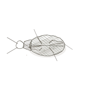
field collected specimen
How to know it on sight
- A thin-walled channel, wider and more irregular than the blood capillaries near it, and often collapsed to a slit.
- No red cells inside. Pale, empty-looking contents, with perhaps a lymphocyte or two.
- A wall one flat cell thick, with no muscle round it and so little basement membrane that the line looks faint or broken.
- In a villus, look in the middle of the core: one pale central channel running the length of it.

Specimen 1

Villi of the jejunum. A lacteal runs up the core of each one. In a routine section it has usually collapsed, so look for a pale slit bounded by a wall one cell thick.
Specimen 2

Villi again, lower power. Blood capillaries lie just under the epithelium; the lacteal sits deeper, in the middle.
Specimen 3

A villus drawn, from Ordo Alimentaria: the lacteal is the single channel up the centre.


People keep calling her by the wrong name
Look-alikes
Two other small vessels with thin walls. Look inside each one.
A blood capillary

A blood capillary — narrower, perfectly round when cut across, with a complete wall. If there is anything inside, it is a red cell.
A venule

A venule — thin-walled and irregular, like a lymphatic. But it holds red cells, and its wall is a continuous line with no gaps in it.
Examine it yourself
The virtual slide
This slide is ileum. Find a villus cut lengthwise, right up its middle. Ignore the epithelium and look at the core. Find the small blood capillaries just under the surface, with red cells in them. Then look deeper, along the centre line, for one wider, paler channel with nothing red in it. That is the lacteal.
Virtual slide courtesy of Histology Guide.
YOur missions
Field Notes for Future Missions
Mission 1: label the capillary
Mission 2: quick check
Field notes
Why this organism
Bladderwort is the plant with no roots that floats just under the surface of still ponds all over the Finger Lakes, and all summer the only part of it you see is a small yellow flower standing clear of the water. Lift a strand and it is hung with hundreds of tiny green sacs. Each sac is hollow and closed, with a door across its mouth. The door is hinged along its top edge and hangs on the inside. Push on it from outside and it swings in. Push on it from inside and it presses harder against its frame. So anything that goes in stays in. That is the wall of a lymphatic capillary. Its cells overlap like that door overlaps its frame: pressure from the tissue side opens the gap, and pressure from the lumen side closes it. Nothing has to decide. The geometry does it. One honest difference. The bladderwort sets its trap by pumping water out of the sac, so that when a bristle is touched the door flies open and the sac sucks. A lymphatic capillary does not suck. It is opened by swelling in the tissue around it, pulling on the filaments that anchor it, and it fills because the pressure outside is, for that moment, higher than the pressure inside.
Next · Pl. II
Hauriens moniliformis var. hormosirae
the string of beads · collecting lymphatic vessel.
In the wild · photograph to come.
Hop to:
← Epithelial Tissues: A Guided Journey
All plates — The Tissue Field Guide: Epithelium
Frontispiece Ordo Epithelia (main page)
Pl. I · Simple squamous
Pl. II · Simple cuboidal
Pl. III · Simple columnar
Pl. IV · Stratified squamous
Pl. V · Stratified cuboidal
Pl. VI · Stratified columnar
Pl. VII · Transitional
Pl. VIII · Pseudostratified columnar
Epithelial Identification Game
All Modules
- Anatomical Language, Membranes & Homeostasis
- Just Enough Chemistry
- The Cell & Its Transport
- Making Cells & Proteins
- The Integumentary System
- The Skeletal System
- The Muscular System
- Nervous Tissue & the Senses
- The Spinal Cord
- The Brain & the Blood–Brain Barrier
- The Autonomic Nervous System
- The Special Senses
- The Endocrine System
- Blood
- The Heart
- Blood Vessels
- The Digestive System
- The Respiratory System
- The Urinary System
- Fluids, Electrolytes & Acid–Base Balance
- The Reproductive System
- The Immune System