Time To Read
Date Last Modified
Pl. II · FAM. CONDUCENTIDAE
Lamellatus ciliatus var. mississippiensis
the plate armor · bronchus
The plate
The specimen, drawn
Pl. II

The specimen as it was drawn, with the wall below it and the lining in the circle.

Habitat & range
Where it lives
Body map

Where this one lives.
Range
- The two main bronchiThe large airways that branch from the trachea into the lungs, dividing into smaller bronchioles. at the carina, then the lobar bronchi — three on the right, two on the left.
- Ten segmental bronchi, one to each bronchopulmonary segment.
- Cartilage plates persist down to roughly a millimetre of diameter, and then stop for good.
- Always inside the lung, and always with a branch of the pulmonary artery running beside it.
Collection record
- Add your slide numbers here.
Niche
What it does for a living
Splits the airstream and keeps distributing it. Still conditioning, still exchanging nothing: a bronchus is plumbing. The complete muscle ring matters because from here downward the airway can change its own diameter, and the cartilageA flexible connective tissue found in joints, the ear, nose, and rib cage. Cartilage can be of three plates are what stop it closing altogether. Every generation of branching doubles the number of tubes and lowers the speed of the air in them, which is why a particle that made it past the trachea has a good chance of settling here.
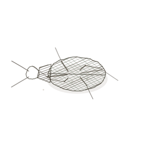
field collected specimen
How to know it on sight
- Cartilage in irregular plates and islands, not a continuous ring. This is the diagnosis.
- A complete ring of smooth muscle has appeared, lying inside the cartilage — the trachea has muscle only across its back.
- Still pseudostratified and ciliated with goblet cellsThe basic structural and functional units of life., but visibly shorter than the trachea’s lining.
- Surrounded by lung. Alveoli on every side, which is what tells you at a glance that you are not in the neckNarrow region just below the head; common fracture site..

Specimen 1

A large airway with lung all around it. Nothing in the trachea’s section A cut or slice of the body or an organ for study. ever looks like this.
Specimen 2

Smaller airways scattered among alveoliMicroscopic air sacs in the lungs where gas exchange occurs between air and blood.. Hunt for cartilage in each one before you name it.
Specimen 3

Hyaline cartilage, here from the trachea — a bronchus’s plates are this same tissue, broken up.


People keep calling her by the wrong name
Look-alikes
Two plates. Read the line underneath each one before you decide.
Trachea

Tell them apart by the cartilage. One ring means trachea; scattered plates mean bronchus. If you can see alveoli in the same field, you are in the lung and it is a bronchus.
Bronchiole

Tell them apart by cartilage, never by size. A large bronchiole is wider than a small bronchus, and sorting by diameter gets this wrong every single time. One island of cartilage is enough to make it a bronchus.
Examine it yourself
The virtual slide
Follow the wall all the way round and mark every piece of cartilage you meet. You are looking for islands with gaps between them — and inside them, a ring of muscle that does not stop.
Virtual slide courtesy of Histology Guide.
YOur missions
Field Notes for Future Missions
Mission 1: label the wall
Mission 2: quick check
Field notes
Why this organism
An alligator’s back is bony plates set in a muscular hide: still armoured, no longer a shell, and now it can bend. That is the step from trachea to bronchus in one image. The armour breaks up into islands at exactly the point the tube needs to be able to move, and the muscle that lets it move closes into a complete ring at the same moment. Two changes, one reason.
Next · Pl. III
Nudus cuboidus var. actinius
the clencher · bronchiole.
In the wild · photograph to come.
Hop to:
← Epithelial Tissues: A Guided Journey
All plates — The Tissue Field Guide: Epithelium
Frontispiece Ordo Epithelia (main page)
Pl. I · Simple squamous
Pl. II · Simple cuboidal
Pl. III · Simple columnar
Pl. IV · Stratified squamous
Pl. V · Stratified cuboidal
Pl. VI · Stratified columnar
Pl. VII · Transitional
Pl. VIII · Pseudostratified columnar
Epithelial Identification Game
All Modules
- Anatomical Language, Membranes & Homeostasis
- Just Enough Chemistry
- The Cell & Its Transport
- Making Cells & Proteins
- The Integumentary System
- The Skeletal System
- The Muscular System
- Nervous Tissue & the Senses
- The Spinal Cord
- The Brain & the Blood–Brain Barrier
- The Autonomic Nervous System
- The Special Senses
- The Endocrine System
- Blood
- The Heart
- Blood Vessels
- The Digestive System
- The Respiratory System
- The Urinary System
- Fluids, Electrolytes & Acid–Base Balance
- The Reproductive System
- The Immune System
List of terms
- bronchi
- cartilage
- cells
- neck
- section
- alveoli