Time To Read
Date Last Modified
Pl. II · FAM. MATURANTIDAE
Maturans stereociliatus
The Long Wait · epididymis
The plate
The specimen, drawn
Pl. II

The worm as it was found, knotted, with the epididymal wall laid out flat below it and the stereocilia read at ×400 in the circle.

Habitat & range
Where it lives
Body map

A single coiled duct lying along the back of each testis, inside the scrotum.
Range
- On the posterior surface of the testis, inside the scrotum — headRounded proximal end that fits into the acetabulum of the hip bone., body and tail, in that order.
- About 5 cm from end to end; uncoiled, the duct runs to roughly six metres.
- Sperm spend somewhere between two days and two weeks in it, and most of that in the tail.
- They arrive unable to swim and leave able to. Nothing else in the tract does that job.
Collection record
- Add your slide numbers here.
Niche
What it does for a living
Finish what the testis started — reabsorb most of the fluid the sperm arrived in, add the secretions that make them motile and able to fertilise, and then hold the finished product until it is called for.
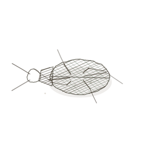
field collected specimen
How to know it on sight
- Roughly circular tubule profiles, close together, each one a section A cut or slice of the body or an organ for study. through the same enormously long duct.
- A tall pseudostratified columnar lining whose nucleiClusters of neurons in the CNS responsible for processing information. sit at two or three different heights, so it looks layered and is not.
- Long tufted projections reaching into the lumenThe inside space of a hollow organ or structure. — stereocilia. They are microvilliTiny projections on the surface of epithelial cells that increase surface area for absorption. and they do not move.
- A thin coat of smooth muscle outside. Thin is the point: compare the wall on Pl. III.

Specimen 1

Tubules in cross-section, the epithelium picked out in darker pink against lighter, mostly clear lumina.
Specimen 2

Testis above, epididymis below, on one slide. The wall changes character completely across that boundary.
Specimen 3

The lining at high power. Pseudostratified columnar, nuclei at several levels, and a dense apical fringe.


People keep calling her by the wrong name
Look-alikes
Two plates, and the first is the single most-missed pair in this volume. The ductus deferens shares this lining exactly; only the wall tells them apart. The second is the gonad upstream, where the cellsThe basic structural and functional units of life. in the wall are the product rather than the plumbing.
Ductus deferens

Same lining family, shorter stereocilia, and a muscularis out of all proportion to the lumen. Muscle-to-lumen ratio decides it.
Seminiferous tubule

A gonad. Four rows of germ cells in the wall itself, getting smaller toward the lumen, and no stereocilia anywhere.
Examine it yourself
The virtual slide
Zoom to about ×10 and centre one lumen. Look at the apical surface first and satisfy yourself the tufts are not ciliaHair-like projections on the surface of some cells that move fluids or particles., then count how many levels the nuclei sit at, then look at how little muscle there is outside.
Virtual slide courtesy of Histology Guide.
YOur missions
Field Notes for Future Missions
Mission 1: find the stereocilia
Mission 2: quick check
Field notes
Why this organism
A horsehair worm spends most of its life as a parasite and emerges as a metre of thread that ties itself into a knot you could hold in your palm. The epididymis is the same joke: six metres of duct packed into something you would not notice. Both solve “I need an enormous length and almost no room” the same way, and both look, to the naked eye, like far less than they are.
Next · Pl. III
Vehens muscularis
The Pinhole Cannon · ductus deferens.
In the wild · photograph to come.
Hop to:
← Epithelial Tissues: A Guided Journey
All plates — The Tissue Field Guide: Epithelium
Frontispiece Ordo Epithelia (main page)
Pl. I · Simple squamous
Pl. II · Simple cuboidal
Pl. III · Simple columnar
Pl. IV · Stratified squamous
Pl. V · Stratified cuboidal
Pl. VI · Stratified columnar
Pl. VII · Transitional
Pl. VIII · Pseudostratified columnar
Epithelial Identification Game
All Modules
- Anatomical Language, Membranes & Homeostasis
- Just Enough Chemistry
- The Cell & Its Transport
- Making Cells & Proteins
- The Integumentary System
- The Skeletal System
- The Muscular System
- Nervous Tissue & the Senses
- The Spinal Cord
- The Brain & the Blood–Brain Barrier
- The Autonomic Nervous System
- The Special Senses
- The Endocrine System
- Blood
- The Heart
- Blood Vessels
- The Digestive System
- The Respiratory System
- The Urinary System
- Fluids, Electrolytes & Acid–Base Balance
- The Reproductive System
- The Immune System
List of terms
- head
- section
- nuclei
- lumen
- microvilli
- cells
- cilia