Time To Read
Date Last Modified
Pl. III · FAM. SIMPLICIDAE
Simplex columnaris var. penicillatus
the brush-bearer · simple columnar epithelium
The plate
The specimen, drawn
Pl. III

The tissue as you will meet it on the slide.

Habitat & range
Where it lives
Body map
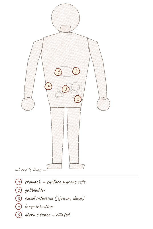
Where this tissue is found.
Range
- Stomach lining — surface mucous cellsThe basic structural and functional units of life.
- Small intestine (jejunumThe middle section of the small intestine, primarily responsible for nutrient absorption., ileumThe last section of the small intestine, responsible for absorbing vitamin B12 and bile salts.)
- Large intestine
- Gallbladder
- Uterine tubes (ciliated)
Collection record
- Slide 029 — simple columnar
- Small intestine, cross-section
- Bronchi slide 130 — ciliated, for comparison
Niche
What it does for a living
Absorption with a surface-area problem to solve. Height gives room for the machinery; microvilliTiny projections on the surface of epithelial cells that increase surface area for absorption. multiply the surface; goblet cells keep it wet. This is the lining of a border crossing that is also a marketplace.
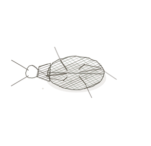
field collected specimen
How to know it on sight
- One row of tall cells standing like a picket fence.
- Nuclei low, near the basement membrane, and all at the same level.
- A brush borderA dense layer of microvilli on the surface of certain cells, increasing surface area for absorption. of microvilli on the free surface.
- Goblet cells punched into the row like white thumbprints.
Specimen 1

Specimen 2

Specimen 3


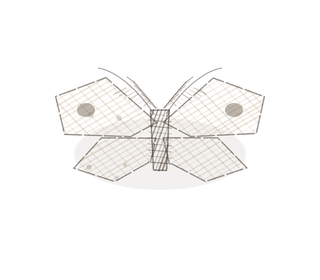
People keep calling her by the wrong name
Look-alikes
Three plates. Read the line underneath each one before you decide.
Pseudostratified columnar

Tell them apart by the nucleiClusters of neurons in the CNS responsible for processing information.. One tidy row = simple. Scattered heights, every cell still touching the membrane = pseudostratified.
Simple cuboidal

Tell them apart by height against width, and where the nucleusThe control center of the cell that contains DNA and directs cellular activities. sits — centred in cuboidal, low in columnar.
Stratified columnar

Tell them apart by looking underneath. A second row of short cells below the tall ones means stratified.
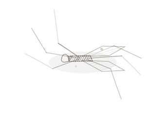
Examine it yourself
The virtual slide
Zoom in until you can see the surface of the cells, then find the basement membrane underneath.
Virtual slide courtesy of Histology Guide.
YOur missions
Field Notes for Future Missions
Mission 1: label the lining
Mission 2: quick check
Field notes
Why this animal
The flamingo’s bill is lined with about a hundred lamellae — an actual brush border — and it feeds with the bill upside down, straining shrimp out of the lake. Tall, narrow, nucleus down by the boots, fuzzy on top.
Next · Pl. IV
Stratifex squamosus var. corneus
the horn-bearer · stratified squamous epithelium.
Hop to:
← Epithelial Tissues: A Guided Journey
All Modules
- Anatomical Language, Membranes & Homeostasis
- Just Enough Chemistry
- The Cell & Its Transport
- Making Cells & Proteins
- The Integumentary System
- The Skeletal System
- The Muscular System
- Nervous Tissue & the Senses
- The Spinal Cord
- The Brain & the Blood–Brain Barrier
- The Autonomic Nervous System
- Special Senses (in developmentThe process of growth and differentiation.)
- The Endocrine System
- Blood
- The Heart
- Blood Vessels
- The Digestive System
- The Respiratory System
- The Urinary System
- Fluids, Electrolytes & Acid–Base Balance
- The Reproductive System
- The Immune System
Plates and body maps drawn for this course. Micrographs from the BIO 171 slide collection. Virtual slides courtesy of Histology Guide.
List of terms
- cells
- jejunum
- ileum
- microvilli
- brush border
- nuclei
- nucleus
- development