Time To Read
Date Last Modified
Pl. IV · FAM. RESPIRANTIDAE
Sacculus squamosus var. riparius
the thin skinSkin found on most of the body, lacking a stratum lucidum. · alveolus
The plate
The specimen, drawn
Pl. IV

The specimen as it was drawn, with the wall below it and the lining in the circle.

Habitat & range
Where it lives
Body map

Where this one lives.
Range
- Roughly 300 million of them, across both lungs.
- About 70 square metres of exchange surface, folded into a chest.
- Alveolar ducts, alveolar sacs, and the alveoliMicroscopic air sacs in the lungs where gas exchange occurs between air and blood. that open off both.
- Present from about week 24 of gestation, and still being added through childhood.
Collection record
- Add your slide numbers here.
Niche
What it does for a living
The only place in this entire volume where gas actually crosses anything. Everything in Plates I to III exists to deliver air to this. The barrier is roughly 0.2 micrometres thick — type I cell, two fused basement membranes, capillary endotheliumThe innermost layer of blood vessels, composed of simple squamous epithelial cells, which reduces fr — and nothing holds an alveolus open except the surface tension of the fluid lining it and the surfactantA substance secreted by Type II pneumocytes that reduces alveolar surface tension. that lowers it. At light-microscope power the wall simply looks thin; if you want to see how thin, Histology Guide’s electron micrograph EM 273 is where to go.
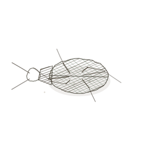
field collected specimen
How to know it on sight
- Thin-walled sacs opening off a shared duct — the grape cluster. At low power the lung looks like lace.
- Type I pneumocytes so flattened that mostly what you see is a nucleusThe control center of the cell that contains DNA and directs cellular activities. bulging from a wall you can barely resolve. They cover about 95 per cent of the surface.
- Type II pneumocytes: domed, cuboidal, bulging into the airspace, usually tucked where two septa meet. These make surfactant.
- Capillaries pressed into the septum with red cellsThe basic structural and functional units of life. sometimes single-file, dust cells loose in the airspace, and pores of Kohn where one septum opens into the next.

Specimen 1

Labelled. Find a flat type I and a domed type II, then read the warning in the look-alikes below.
Specimen 2

The lace at low power. Every pale line is a septum with a capillary inside it.
Specimen 3

Alveolar sacs opening off a duct. Follow one sac back to the duct it belongs to.


People keep calling her by the wrong name
Look-alikes
Two plates. Read the line underneath each one before you decide.
Pneumonia

Tell them apart by what is in the airspace. Healthy alveoli are empty; here they are packed with exudate and cells. Worth seeing on purpose, because the architecture is intact and the function is gone.
Respiratory bronchiole

Tell them apart by whether there is any wall. A respiratory bronchiole keeps scraps of cuboidal lining between the alveolar mouths; an alveolar duct is nothing but mouths with knobs of muscle between them.
Examine it yourself
The virtual slide
Hunt for a type II cell. It is domed and it bulges, and it is usually sitting in the corner where two septa meet. Then check Histology Guide’s own label on it — and read the warning below before you copy it down.
Virtual slide courtesy of Histology Guide.
YOur missions
Field Notes for Future Missions
Mission 1: label the wall
Mission 2: quick check
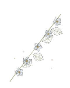
Field notes
Why this organism
A grape cluster is thin-skinned sacs on a shared stalk, which is the alveolar sac and its duct. The anatomists got there first: acinus is Latin for berry, and the pulmonary acinus is still what the unit is called. What the grape carries best is that the skinThe body’s largest organ, providing protection and regulation. is all there is. One warning about the slide itself: on both MH 138 and MH 139, Histology Guide labels the domed cuboidal cell Type I. That is the type II cell. Flat and nearly invisible is type I; domed and bulging is type II. Two different slides carry the same error, so it is not a one-off.
Next · Pl. V
Operculum stratificatus var. dionaeus
the trapdoor · epiglottisA flap of cartilage that covers the trachea during swallowing to prevent food from entering the airw.
In the wild · photograph to come.
Hop to:
← Epithelial Tissues: A Guided Journey
All plates — The Tissue Field Guide: Epithelium
Frontispiece Ordo Epithelia (main page)
Pl. I · Simple squamous
Pl. II · Simple cuboidal
Pl. III · Simple columnar
Pl. IV · Stratified squamous
Pl. V · Stratified cuboidal
Pl. VI · Stratified columnar
Pl. VII · Transitional
Pl. VIII · Pseudostratified columnar
Epithelial Identification Game
All Modules
- Anatomical Language, Membranes & Homeostasis
- Just Enough Chemistry
- The Cell & Its Transport
- Making Cells & Proteins
- The Integumentary System
- The Skeletal System
- The Muscular System
- Nervous Tissue & the Senses
- The Spinal Cord
- The Brain & the Blood–Brain Barrier
- The Autonomic Nervous System
- The Special Senses
- The Endocrine System
- Blood
- The Heart
- Blood Vessels
- The Digestive System
- The Respiratory System
- The Urinary System
- Fluids, Electrolytes & Acid–Base Balance
- The Reproductive System
- The Immune System
List of terms
- thin skin
- alveoli
- endothelium
- surfactant
- nucleus
- cells
- skin
- epiglottis