Time To Read
Date Last Modified
Pl. IV · FAM. IUNCTIDAE
Iunctio neuralis var. electrophorus
the private line · the neuromuscular junction
The plate
The specimen, drawn
Pl. IV

The junction as you will meet it on the slide.

Habitat & range
Where it lives
Body map

Where this junction is found.
Range
- One on nearly every skeletal muscle fiber you have, and with few exceptions exactly one. The exceptions are worth knowing: the fibers inside a muscle spindle, and about a fifth of the fibers in the eye muscles, carry several endings each.
- Usually near the middle of the fiber, so the impulse has the same distance to travel to each end of it.
- The six muscles that aim each eye, where one neuron may own only three or four fibers — the smallest motor units in the body, and the first to show when the junction is in trouble.
- The diaphragm, about twelve times a minute, whether or not you are paying any attention to it.
- Smooth muscle has nothing like this. The autonomic axon runs past the cellsThe basic structural and functional units of life. with swellings strung along it and lets the transmitter find its own way, which is the comparison this whole plate is built around.
Collection record
- Add your slide numbers here.
Niche
What it does for a living
It is a relay with one job: turn a nerve impulse into a muscle impulse, and never drop one. The axon terminalThe endpoint of an axon where neurotransmitters are stored and released into a synapse. releases acetylcholinealso know as ACh A neurotransmitter that stimulates muscle contraction. into a gap about fifty nanometres across; the muscle membrane underneath is thrown into deepAway from the surface of the body. folds packed with receptorsProteins located on the surface or inside cells that bind specific molecules (e.g., neurotransmitter; and the end plate potential that follows is several times larger than it needs to be. That deliberate excess is the safety factor, and it is why the junction is the quietest link in the whole chain — one impulse in, one twitch out, every single time. It only becomes interesting when something eats into the margin. Myasthenia gravis destroys the receptors, curare blocks them, and organophosphates jam the enzyme that clears the transmitter away. In each case the failure shows up first where the margin was thinnest to begin with: the eye muscles have shallower junctional folds and fewer receptors packed into them than the rest of you do, which is why the eyelids and the eyes are usually what goes first.
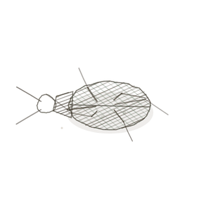
field collected specimen
How to know it on sight
- One dark, branching smudge sitting on the surface of a fiber — on it, not inside it, and not between fibers.
- Once per fiber. If you are seeing a mark on every fiber in the field, you are looking at nucleiClusters of neurons in the CNS responsible for processing information., not junctions.
- The nerve arrives as a single thin fibre that divides only at the very end, and loses its myelin sheathA fatty covering around axons that increases conduction speed. just before it does.
- It will not show on an ordinary H and E section A cut or slice of the body or an organ for study.. Junctions need a stain that picks out nerve — gold chloride, silver, or a label for the receptors.

Specimen 1

Gold chloride blackens nerve and leaves muscle pale. Find the thin dark line crossing the field, follow it to where it stops, and that spray of branches is the terminal — spread flat on the fiber, never buried in it.
Specimen 2

The same preparation, another field. Count how many fibers carry a terminal. Most do not, because the fibers belonging to one motor unitA motor neuron and all the muscle fibers it controls. are scattered through the muscle rather than lined up side by side.
Specimen 3

Ordinary skeletal muscle, H and E, and not a junction in sight — which is the point. Every one of these fibers has one. The dark ovals you can see are nuclei pressed against the rim, and they are not it.


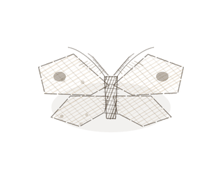
People keep calling her by the wrong name
Look-alikes
Two plates. Read the line underneath each one before you decide.
A connective tissue nucleus

Tell them apart by which side of the membrane it is on. The nuclei scattered between the fibers belong to the endomysium — fibroblasts, capillary cells, the occasional wandering immune cell. A junction sits on a fiber, is much larger than any nucleusThe control center of the cell that contains DNA and directs cellular activities., and has a nerve leading to it that you can follow.
Smooth muscle and its varicosities

Tell them apart by looking for an address. Smooth muscle is reached by an axon that passes among the cells with beads strung along it, releasing transmitter into the shared space between them. No terminal, no end plate, no folds — and no one cell the message was ever meant for.
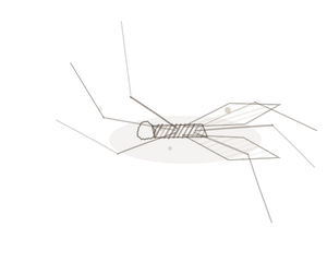
Examine it yourself
The virtual slide
This one is an electron micrograph rather than a light slide, because the part that matters most is below the resolution of your microscope. Find the axon terminal, then the gap, then the folds in the muscle membrane underneath it — and work out roughly how much extra membrane those folds buy.
Electron micrograph courtesy of Histology Guide.
YOur missions
Field Notes for Future Missions
Mission 1: find the junction
Mission 2: quick check
Field notes
Why this organism
An electric eel is four fifths battery. The organ runs almost the whole length of the body, and every plate in it — around six thousand of them, stacked headRounded proximal end that fits into the acetabulum of the hip bone. to tail — is a muscle cell that gave up contracting and kept the junction. Each electrocyte is innervated on one face only, never on both, and because the nerve supply reaches all of them in the same instant the small voltages add instead of cancelling. That is the neuromuscular junction with the muscle part taken away: a dedicated line, one address, one message, and no room for a plate to decide anything on its own. The shock it makes does the opposite thing — it spreads out into the waterThe universal solvent essential for life. and acts on whatever happens to be standing there, which is very nearly what an autonomic axon does to a sheet of smooth muscle.
Next · Pl. V
Iunctio tendinea var. mytilus
the splice · the myotendinous junction.
In the wild
The organism, photographed

Pexels has no photograph of Electrophorus, so here is the water instead — a blackwater tributary of the Amazon, tea-dark and nearly lightless below the surface. An animal here does not find its dinner by looking at it. Photograph: nandofreitasr / Pexels.
Hop to:
← Epithelial Tissues: A Guided Journey
All plates — The Tissue Field Guide: Epithelium
Frontispiece Ordo Epithelia (main page)
Pl. I · Simple squamous
Pl. II · Simple cuboidal
Pl. III · Simple columnar
Pl. IV · Stratified squamous
Pl. V · Stratified cuboidal
Pl. VI · Stratified columnar
Pl. VII · Transitional
Pl. VIII · Pseudostratified columnar
Epithelial Identification Game
All Modules
- Anatomical Language, Membranes & Homeostasis
- Just Enough Chemistry
- The Cell & Its Transport
- Making Cells & Proteins
- The Integumentary System
- The Skeletal System
- The Muscular System
- Nervous Tissue & the Senses
- The Spinal Cord
- The Brain & the Blood–Brain Barrier
- The Autonomic Nervous System
- The Special Senses
- The Endocrine System
- Blood
- The Heart
- Blood Vessels
- The Digestive System
- The Respiratory System
- The Urinary System
- Fluids, Electrolytes & Acid–Base Balance
- The Reproductive System
- The Immune System
List of terms
- neuromuscular junction
- cells
- axon terminal
- acetylcholine
- deep
- receptors
- nuclei
- myelin sheath
- section
- motor unit
- nucleus
- head
- water