Time To Read
Date Last Modified
Pl. IV · FAM. STRATIFICIDAE
Stratifex squamosus var. corneus
the horn-bearer · stratified squamous epithelium
The plate
The specimen, drawn
Pl. IV

The tissue as you will meet it on the slide.

Habitat & range
Where it lives
Body map
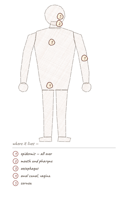
Where this tissue is found.
Range
- Epidermis
- Oral cavity, pharynxThe muscular passageway connecting the mouth to the esophagus and larynx., oesophagus
- Anal canal
- Vagina
- Cornea
Collection record
- Add your slide numbers here.
Niche
What it does for a living
Wear and replacement. It expects to lose its surface and is built to keep replacing it from below. Protection, not exchange — nothing useful crosses this tissue quickly, which is exactly the point.
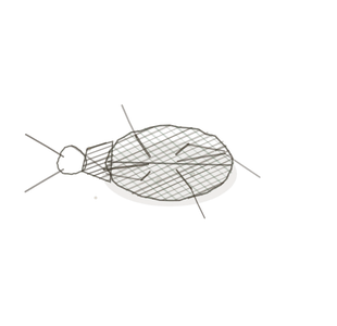
field collected specimen
How to know it on sight
- Many layers, and the cellsThe basic structural and functional units of life. change shape as you climb.
- Cuboidal and busy at the bottom, polygonal in the middle, flat and dying on top.
- Named for the top layer, not the bottom.
- Keratinised where it is dry; still nucleated where it is wet.
Specimen 1

Specimen 2

Specimen 3


People keep calling her by the wrong name
Look-alikes
Three plates. Read the line underneath each one before you decide.
Transitional

Tell them apart by the top row and the address. Transitional domes rather than flattens, and lives only in the urinary tract.
Stratified cuboidal

Tell them apart by depth. Two layers with square top cells, not a thick stack of flattening ones.
Pseudostratified

Tell them apart by tracing cells down. If every one reaches the membrane it was never stratified.
Examine it yourself
The virtual slide
Zoom in until you can see the surface of the cells, then find the basement membrane underneath.
Virtual slide courtesy of Histology Guide.
YOur missions
Field Notes for Future Missions
Mission 1: label the lining
H5P image hotspots — paste the shortcode here.
Mission 2: quick check
Field notes
Why this animal
A pangolin’s scales are keratinA strong, fibrous protein that forms the structure of skin, hair, and nails., laid down at the baseA substance that accepts hydrogen ions (H⁺) or releases hydroxide ions (OH⁻). and worn away at the tip. It is wearing the tissue on the outside.
Next · Pl. V
Stratifex cubicus var. geminus
the twinned · stratified cuboidal epithelium.
Hop to:
← Epithelial Tissues: A Guided Journey
All Modules
- Anatomical Language, Membranes & Homeostasis
- Just Enough Chemistry
- The Cell & Its Transport
- Making Cells & Proteins
- The Integumentary System
- The Skeletal System
- The Muscular System
- Nervous Tissue & the Senses
- The Spinal Cord
- The Brain & the Blood–Brain Barrier
- The Autonomic Nervous System
- Special Senses (in developmentThe process of growth and differentiation.)
- The Endocrine System
- Blood
- The Heart
- Blood Vessels
- The Digestive System
- The Respiratory System
- The Urinary System
- Fluids, Electrolytes & Acid–Base Balance
- The Reproductive System
- The Immune System
Plates and body maps drawn for this course. Micrographs from the BIO 171 slide collection. Virtual slides courtesy of Histology Guide.
List of terms
- pharynx
- cells
- keratin
- base
- development