Time To Read
Date Last Modified
Pl. IX · FAM. VEHENTIDAE
Vehens ciliatus
The Feather Conveyor · uterine tube


Habitat & range
Where it lives

Range
- Paired, running laterally from the upper corners of the uterus in the free edge of the broad ligament.
- About 10 cm long: infundibulumThe stalk connecting the hypothalamus to the pituitary gland. with its fimbriae, then ampulla, then isthmusA narrow portion of the uterine tube connecting to the uterus., then the intramural part.
- The fimbriae are not attached to the ovary. The oocyte crosses a gap to be caught.
- Fertilisation normally happens in the ampulla, the widest and most folded part.
Collection record
- Add your slide numbers here.
Niche
What it does for a living
Catch an oocyte that was never connected to anything, host fertilisation, and deliver the result to the uterus on a schedule the endometriumThe inner lining of the uterus that thickens during the menstrual cycle to support a potential pregn is keeping independently.
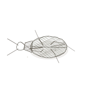
field collected specimen
How to know it on sight
- A mucosaThe innermost lining of the digestive tract that contains mucus-secreting cells for protection and a thrown into deepAway from the surface of the body. branching folds that very nearly fill the lumenThe inside space of a hollow organ or structure. — labyrinthine, especially in the ampulla.
- A simple columnar lining, one row of nucleiClusters of neurons in the CNS responsible for processing information. at much the same level. Not pseudostratified.
- True ciliaHair-like projections on the surface of some cells that move fluids or particles.: even, upright, and standing on a dark line of basal bodies.
- Between them, rounded non-ciliated peg cellsThe basic structural and functional units of life. bulging above the ciliated surface, which is why the apex looks uneven.

Specimen 1
The tube in cross-section. Concentric layers around an irregular central space nearly closed by folds.
Specimen 2

Her labelled tract diagram, for orientation: where the tube runs and how the fimbriae relate to the ovary.
Specimen 3

The ovary the fimbriae sweep. The oocyte released from a follicle like this is what the cilia have to catch.


People keep calling her by the wrong name
Look-alikes
Two plates. The first has an apical fringe that looks similar at low power and is not cilia at all; proving that to yourself is the point of this plate. The second is the organ downstream, which shares the lining family but has no folds like these.
Epididymis

Tufted, leaning, uneven, and pseudostratified beneath. Cilia are even, upright, and sit on a line of basal bodies.
Uterus

Simple columnar again, but the mucosa makes tubular glands that dive into stroma rather than folds that fill the lumen.
Examine it yourself
The virtual slide
Zoom to ×20 and centre the lining. Look along the apical surface for the dark line of basal bodies, then find a cell that bulges above its neighbours and has no cilia on it.
Virtual slide courtesy of Histology Guide.
YOur missions
Field Notes for Future Missions
Mission 1: cilia or stereocilia
Mission 2: quick check
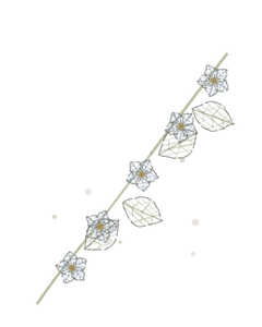
Field notes
Why this organism
A feather star holds its arms up into the current and runs food down a ciliated groove on each one to the mouthThe opening of the digestive tract where food enters and mastication begins. at the centre. It does not chase anything; it makes the waterThe universal solvent essential for life. do the work. The uterine tube’s fimbriae sweep over an ovary they are not attached to and catch an oocyte with cilia, and the oocyte has no means of propulsion of its own. If this tube were lined with stratified squamous instead, there would be no cilia, nothing would move, and an embryo would implant where it sat — which is what an ectopic pregnancy is.
Next · Pl. X
Recipiens cyclicus
The Sap Run · uterus.
In the wild · photograph to come.
Hop to:
← Epithelial Tissues: A Guided Journey
All plates — The Tissue Field Guide: Epithelium
Frontispiece Ordo Epithelia (main page)
Pl. I · Simple squamous
Pl. II · Simple cuboidal
Pl. III · Simple columnar
Pl. IV · Stratified squamous
Pl. V · Stratified cuboidal
Pl. VI · Stratified columnar
Pl. VII · Transitional
Pl. VIII · Pseudostratified columnar
Epithelial Identification Game
All Modules
- Anatomical Language, Membranes & Homeostasis
- Just Enough Chemistry
- The Cell & Its Transport
- Making Cells & Proteins
- The Integumentary System
- The Skeletal System
- The Muscular System
- Nervous Tissue & the Senses
- The Spinal Cord
- The Brain & the Blood–Brain Barrier
- The Autonomic Nervous System
- The Special Senses
- The Endocrine System
- Blood
- The Heart
- Blood Vessels
- The Digestive System
- The Respiratory System
- The Urinary System
- Fluids, Electrolytes & Acid–Base Balance
- The Reproductive System
- The Immune System
List of terms
- current
- section
- uterus
- fimbriae
- infundibulum
- isthmus
- endometrium
- mucosa
- deep
- lumen
- nuclei
- cilia
- cells
- mouth
- water