Time To Read
Date Last Modified
Pl. V · FAM. GLANDULIDAE
Glandula labyrinthica
The False Rooms · seminal vesicle
The plate
The specimen, drawn
Pl. V

The fungus as it was found, with the folded wall below it and the fold hierarchy read at ×200 in the circle.

Habitat & range
Where it lives
Body map

Paired, behind the bladderA muscular organ that stores urine before excretion., joining the ductus deferens above the prostate.
Range
- Paired, on the posterior surface of the bladder, above the prostate and in front of the rectumThe final section of the large intestine, where feces are stored before elimination..
- About 5 cm long as an organ — but it is one coiled tube roughly 15 cm long when unwound.
- Its duct joins the ampulla of the ductus deferens to form the ejaculatory duct.
- Supplies about 60% of semen by volume: fructose-rich, alkaline, and yellowish.
Collection record
- Add your slide numbers here.
Niche
What it does for a living
Supply the bulk, the fuel and the pHA measure of hydrogen ion concentration in a solution. — fructose to power the flagellum, and alkali to buffer an environment that would otherwise immobilise everything within minutes.
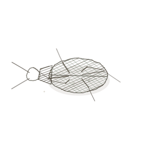
field collected specimen
How to know it on sight
- A honeycomb of apparent chambers, all of them continuous with one another. Trace the lumenThe inside space of a hollow organ or structure. and it is one space.
- Primary folds thrown off the wall, secondary folds off those, tertiary off those again.
- A tall simple to pseudostratified columnar lining, secretory, often with pale secretionThe process of moving substances from the blood into the nephron tubule to be excreted in urine. in the lumen.
- A modest muscular coat outside. This is a storage bag, not a pump.

Specimen 1

The folded architecture at low power. Count the chambers, then try to count them again from a different starting point.
Specimen 3

The spermatic cord for contrast: genuinely separate structures, against the single folded space here.


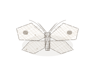
People keep calling her by the wrong name
Look-alikes
Two plates. The first is the other large accessory gland, and the architecture separates them. The second is a duct whose profiles really are separate, which is the comparison that makes the point about this one.
Prostate

Irregular pockets of every size, genuinely separate from one another, set into smooth muscle, and sometimes holding a stone.
Epididymis

Here the profiles are separate: each is a section A cut or slice of the body or an organ for study. through a coiled duct. In the seminal vesicle they only look that way.
Examine it yourself
The virtual slide
Click Mucosa and go to ×40. Pick one apparent chamber and follow its wall with the cursor until it opens into the next one. Do that twice and the organ stops looking like a honeycomb.
Virtual slide courtesy of Histology Guide.
YOur missions
Field Notes for Future Missions
Mission 1: trace one lumen
Mission 2: quick check
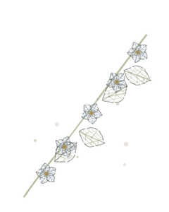
Field notes
Why this organism
Clathrus ruber fruits as a hollow red lattice — a ball of branching arms with gaps between them. Look at a slice of it and you will count a dozen chambers that are all the same single interior. That is the one mistake every student makes on this slide: counting rooms in a building that has one room. Tracing the lumen is the whole skill, and a stinkhorn makes the error visible from the outside.
Next · Pl. VI
Glandula lubricans
The Slipstream · bulbourethral gland.
In the wild · photograph to come.
Hop to:
← Epithelial Tissues: A Guided Journey
All plates — The Tissue Field Guide: Epithelium
Frontispiece Ordo Epithelia (main page)
Pl. I · Simple squamous
Pl. II · Simple cuboidal
Pl. III · Simple columnar
Pl. IV · Stratified squamous
Pl. V · Stratified cuboidal
Pl. VI · Stratified columnar
Pl. VII · Transitional
Pl. VIII · Pseudostratified columnar
Epithelial Identification Game
All Modules
- Anatomical Language, Membranes & Homeostasis
- Just Enough Chemistry
- The Cell & Its Transport
- Making Cells & Proteins
- The Integumentary System
- The Skeletal System
- The Muscular System
- Nervous Tissue & the Senses
- The Spinal Cord
- The Brain & the Blood–Brain Barrier
- The Autonomic Nervous System
- The Special Senses
- The Endocrine System
- Blood
- The Heart
- Blood Vessels
- The Digestive System
- The Respiratory System
- The Urinary System
- Fluids, Electrolytes & Acid–Base Balance
- The Reproductive System
- The Immune System
List of terms
- bladder
- rectum
- pH
- lumen
- secretion
- cells
- nuclei
- base
- section