Time To Read
Date Last Modified
Pl. VII · FAM. VIGILIDAE
Vigilans appendicis var. abelmoschi
the dead end · appendix
The plate
The specimen, drawn
Pl. VII

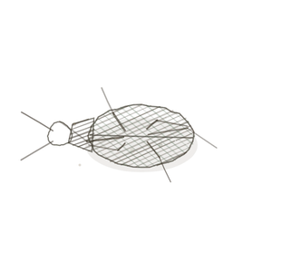
The okra pod as it was drawn, whole and cut across, with the appendix in cross-section below it and one of its follicles in the circle.

Habitat & range
Where it lives
Body map

Off the cecum, in the right lower abdomen, at the very start of the large intestine.
Range
- A blind tube about as long as a finger, opening off the cecum just below where the small intestine joins.
- Its tip can lie almost anywhere nearby, which is why the pain of appendicitis is not always where the textbook puts it.
- It is one outpost of the gut’s lymphoid tissue. The other two, the tonsils and Peyer’s patches, are on Ordo Alimentaria, Pl. XI.
- All three are MALT, mucosa-associated lymphoid tissue: follicles built into the wall of a tube, with no capsule round them.
Collection record
- Add your slide numbers here.
Niche
What it does for a living
Stand guard in the wall of the gut, at a dead end where the contents sit still. The appendix has the four layers of any piece of the digestive tube: a mucosa, a submucosa, a muscle coat and a serosa. What sets it apart is how much of the wall is given over to lymphoid tissue. Follicles fill the mucosa and spill into the submucosa, and in a young person they form a complete ring round the lumen, shoulder to shoulder. Many have germinal centres, because they are in permanent contact with what the gut contains. The epithelium over each follicle samples the contents and hands fragments down to the lymphocytes beneath, and B cells primed here go on to make the antibody that protects the whole lining of the gut. So this is the same kind of tissue as a Peyer’s patch or a tonsil (Ordo Alimentaria, Pl. XI), and it differs from a lymph node (Pl. III) in one important way: there is no capsule and there are no afferent lymphatics. Nothing is delivered. The tissue sits directly under the surface it watches. The lumen is narrow, and that is the organ’s weakness. If it blocks, the appendix swells behind the blockage, its blood supply fails, and it can burst.
field collected specimen
How to know it on sight
- A small, complete cross-section of gut: round, with a narrow, irregular lumen and often debris in it.
- A ring of lymphoid follicles in the wall, right round the lumen, some with pale germinal centres. This is the field mark.
- Short, sparse crypts and no villi. The lining is like the colon’s, but there is far less of it.
- No capsule round the lymphoid tissue. Outside it are a submucosa, a complete coat of smooth muscle, and a serosa.

Specimen 1

Where it sits: at the lower right of the abdomen, off the first part of the large intestine.
Specimen 2

A Peyer’s patch, drawn, from Ordo Alimentaria. The same follicles under the same kind of dome, in the wall of the ileum.
Specimen 3

A tonsil. Follicles under an epithelium again, but here the epithelium is stratified squamous and dips into deep crypts.


People keep calling her by the wrong name
Look-alikes
Two other cross-sections of gut. Count the follicles, and look for villi.
Colon

Colon — the same lining, with straight crypts and no villi, but a wide lumen and only the occasional follicle. Nothing like a complete ring.
Small intestine

Small intestine — villi, which the appendix never has. Follicles here are gathered into Peyer’s patches on one side of the wall only.
Examine it yourself
The virtual slide
At the lowest power the whole organ should fit in the field. Find the lumen, then count the follicles round it. Pick one with a pale centre and go up on it. Then work outward: find where the lymphoid tissue ends, and name each layer between there and the outside.
Virtual slide courtesy of Histology Guide.
YOur missions
Field Notes for Future Missions
Mission 1: label the wall
Mission 2: quick check
Field notes
Why this organism
Okra comes to the farm stands on Route 53 at the end of August, and anyone who has cooked it knows two things about it. The pod is a narrow, ridged finger that tapers to a closed tip: it hangs off the plant by one end and goes nowhere at the other. And when you slice it across, every slice is the same wheel. A ring of wall, a small hollow in the middle, and round seeds set at even intervals all the way round. The appendix is built to that picture. It is a finger-shaped pouch hanging off the cecum, open at one end and closed at the other. Cut it across and you find a small lumen in the middle and a ring of round lymphoid follicles in the wall, evenly spaced, the whole way round. No other piece of gut looks like that in cross-section. The colon has a follicle here and there, and the ileum gathers them into patches on one side. One honest difference. Okra seeds are the reason the pod exists, and each sits in its own compartment. The follicles of the appendix are not sealed off in chambers. They lie in ordinary connective tissue with no capsule round them, lymphocytes drifting between one and the next, directly under the lining they are there to watch. And the count changes: the ring is fullest in a teenager and thins with every decade after. For the other two places the gut keeps follicles like these, the tonsils at its entrance and the Peyer’s patches of the ileum, see Ordo Alimentaria, Pl. XI.
Back · Frontispiece
Ordo Custodia
the sentries · the whole volume.
In the wild · photograph to come.
Hop to:
← Epithelial Tissues: A Guided Journey
All plates — The Tissue Field Guide: Epithelium
Frontispiece Ordo Epithelia (main page)
Pl. I · Simple squamous
Pl. II · Simple cuboidal
Pl. III · Simple columnar
Pl. IV · Stratified squamous
Pl. V · Stratified cuboidal
Pl. VI · Stratified columnar
Pl. VII · Transitional
Pl. VIII · Pseudostratified columnar
Epithelial Identification Game
All Modules
- Anatomical Language, Membranes & Homeostasis
- Just Enough Chemistry
- The Cell & Its Transport
- Making Cells & Proteins
- The Integumentary System
- The Skeletal System
- The Muscular System
- Nervous Tissue & the Senses
- The Spinal Cord
- The Brain & the Blood–Brain Barrier
- The Autonomic Nervous System
- The Special Senses
- The Endocrine System
- Blood
- The Heart
- Blood Vessels
- The Digestive System
- The Respiratory System
- The Urinary System
- Fluids, Electrolytes & Acid–Base Balance
- The Reproductive System
- The Immune System