Time To Read
Date Last Modified
Pl. VIII · FAM. SUSTINENTIDAE
Sustinens elasticus var. auritus
the bouncer · elastic cartilageA flexible connective tissue found in joints, the ear, nose, and rib cage. Cartilage can be of three
The plate
The specimen, drawn
Pl. VIII

Hyaline with a dark elastic mesh woven through it. Hold the shape, and bend without complaint.

Habitat & range
Where it lives
Body map

Only three places in the whole body, and that really is the list.
Range
- The external ear — fold it and let go.
- The epiglottisA flap of cartilage that covers the trachea during swallowing to prevent food from entering the airw, which has to fold over the airway and spring back.
- The auditory tube, and the corniculate and cuneiform cartilages of the larynxThe voice box; contains vocal cords and connects the pharynx to the trachea..
Collection record
- Add your slide numbers here.
Niche
What it does for a living
Keeping a shape that has to be deformed regularly. The ear gets slept on, knocked and folded every day of your life and returns to the same shape each time; the epiglottis does the same several times a minute. Hyaline would crack and dense regular would not hold a shape at all, so this tissue takes the cartilage framework and threads elastin through it.
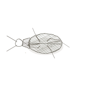
field collected specimen
How to know it on sight
- ChondrocytesMature cartilage cells housed in lacunae. in lacunae, as in hyaline — the cellsThe basic structural and functional units of life. look the same.
- But the matrix is full of a dark branching network of elastic fibres.
- The network is densest in the middle and thins towards the perichondrium.
- Needs an elastic stain to be obvious; in plain H&E it can pass for hyaline.

Specimen 1

Masson trichrome. The chondrocytes are clear; look at how much texture there is in the matrix between them.

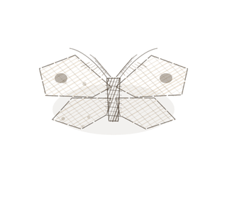
People keep calling her by the wrong name
Look-alikes
Two plates. Read the line underneath each one before you decide.
Hyaline cartilage

Same cells, same lacunae. The question is only whether the matrix is featureless or has a fibre network in it.
Fibrocartilage

Fibrocartilage bundles are thick, pink and parallel; elastic fibres are fine, dark and branching.
Examine it yourself
The virtual slide
Travel from the centre of the cartilage out to the perichondrium and watch the elastic network thin out as you go. Then ask why the middle would need more of it than the edge.
Virtual slide courtesy of Histology Guide.
YOur missions
Field Notes for Future Missions
Mission 1: the hidden mesh
Mission 2: quick check
Field notes
Why this organism
A fennec fox has ears nearly a third of its body length, and it sleeps on them, pushes through burrows with them and swivels them constantly, and they have never once needed re-setting. The ear is thin enough to shed heat and springy enough to survive being folded flat. That combination — a shape that is held, but held flexibly — is the one problem elastic cartilage exists to solve, and the body only found three places that needed it.
Next · Pl. IX
Sustinens fibrosus var. nuciferus
the shock absorber · fibrocartilageA tough, shock-absorbing cartilage found in intervertebral discs and menisci..
Hop to:
← Epithelial Tissues: A Guided Journey
All plates — The Tissue Field Guide: Epithelium
Frontispiece Ordo Epithelia (main page)
Pl. I · Simple squamous
Pl. II · Simple cuboidal
Pl. III · Simple columnar
Pl. IV · Stratified squamous
Pl. V · Stratified cuboidal
Pl. VI · Stratified columnar
Pl. VII · Transitional
Pl. VIII · Pseudostratified columnar
Epithelial Identification Game
All Modules
- Anatomical Language, Membranes & Homeostasis
- Just Enough Chemistry
- The Cell & Its Transport
- Making Cells & Proteins
- The Integumentary System
- The Skeletal System
- The Muscular System
- Nervous Tissue & the Senses
- The Spinal Cord
- The Brain & the Blood–Brain Barrier
- The Autonomic Nervous System
- The Special Senses
- The Endocrine System
- Blood
- The Heart
- Blood Vessels
- The Digestive System
- The Respiratory System
- The Urinary System
- Fluids, Electrolytes & Acid–Base Balance
- The Reproductive System
- The Immune System
List of terms
- elastic cartilage
- cartilage
- epiglottis
- larynx
- Chondrocytes
- cells
- fibrocartilage