Time To Read
Date Last Modified
Pl. VIII · FAM. GENERANTIDAE
Generans luteus
The Afterglow · corpus luteum
The plate
The specimen, drawn
Pl. VIII

The same plant seven days on, with the luteum and the albicans set side by side below it and the folded wall read at ×40 in the circle.

Habitat & range
Where it lives
Body map

The same ovary as Pl. VII. This plate is a time, not a place.
Range
- In the ovarian cortex, at the site where a follicle ruptured — so its position is wherever ovulationThe release of a mature oocyte from the ovary. happened.
- Up to 2 cm across at its peak, which makes it the largest single structure in a normal ovary.
- It lasts about fourteen days without pregnancy, and several months with one.
- Old corpora albicantia accumulate; an ovary late in reproductive life is full of them.
Collection record
- Add your slide numbers here.
Niche
What it does for a living
Be a gland that is built to die on schedule. It makes the progesteroneA hormone that supports pregnancy and regulates the menstrual cycle. that holds the endometriumThe inner lining of the uterus that thickens during the menstrual cycle to support a potential pregn, and the withdrawal of that progesterone when it degenerates is itself the signal that starts menstruation.
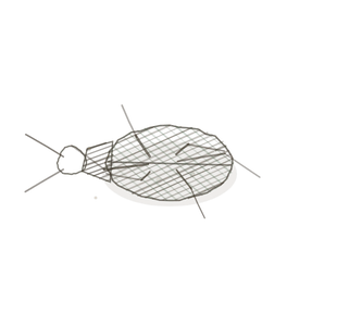
field collected specimen
How to know it on sight
- A large, pale, conspicuously lobulated mass — the wall is thrown into deepAway from the surface of the body. folds because the follicle collapsed.
- Large pale cellsThe basic structural and functional units of life. with vesicular nucleiClusters of neurons in the CNS responsible for processing information. filling the folds: granulosa-lutein, with smaller theca-lutein cells at the edges.
- No antrum and no oocyte. If either is still there, you are looking at a follicle, not a corpus luteum.
- Nearby, one or more small dense white bodies with no cells worth the name: corpora albicantia, from earlier cycles.

Specimen 1

Granulosa and thecaA layer of cells surrounding the follicle that produces hormones. lutein cellsCells in the corpus luteum that produce hormones. at higher power, and the vascularity that arrives when the follicle ruptures.
Specimen 2

The whole ovary. Find the corpus luteum first by size alone, then hunt the small dense scars around it.
Specimen 3

Tertiary folliclesStructures in the ovaries that contain developing oocytes. for contrast — antrum present, oocyte present, wall not folded.


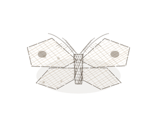
People keep calling her by the wrong name
Look-alikes
Two plates. The first is the stage immediately before, and the presence of an antrum settles it. The second is the look-alike that actually costs marks: another pale, lipid-rich, corded endocrine tissue that students meet in the same semester.
Tertiary follicle
Before the event. A fluid-filled antrum and an oocyte in its zona pellucidaA glycoprotein layer surrounding the oocyte.; the wall is a smooth ring, not a folded one.
Ovary, whole
The context. At this magnification the corpus luteum is unmistakable on size, and the albicantia show as small dense bodiesStructures in smooth muscle that anchor actin filaments..
Examine it yourself
The virtual slide
Without magnifying, find the biggest thing in the section A cut or slice of the body or an organ for study.. Check it has no antrum and no oocyte. Then look around it for at least two small, dense, pale scars.
Virtual slide courtesy of Histology Guide.
YOur missions
Field Notes for Future Missions
Mission 1: luteum or albicans
Mission 2: quick check
Field notes
Why this organism
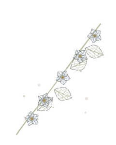
Pl. VII is the same plant one week later. A lily that opened and closed and sank leaves a swollen seed capsule riding just under the surface, and then a pale fibrous knot. It is the only mascot in the guide that shares an organismA living individual made up of cells. with its neighbour on purpose — the second time the roster has done that, after the stony coral on Conjunctiva Pl. X and XI, and for the same reason: these are not two things, they are one thing at two times.
Next · Pl. IX
Vehens ciliatus
The Feather Conveyor · uterine tube.
In the wild · photograph to come.
Hop to:
← Epithelial Tissues: A Guided Journey
All plates — The Tissue Field Guide: Epithelium
Frontispiece Ordo Epithelia (main page)
Pl. I · Simple squamous
Pl. II · Simple cuboidal
Pl. III · Simple columnar
Pl. IV · Stratified squamous
Pl. V · Stratified cuboidal
Pl. VI · Stratified columnar
Pl. VII · Transitional
Pl. VIII · Pseudostratified columnar
Epithelial Identification Game
All Modules
- Anatomical Language, Membranes & Homeostasis
- Just Enough Chemistry
- The Cell & Its Transport
- Making Cells & Proteins
- The Integumentary System
- The Skeletal System
- The Muscular System
- Nervous Tissue & the Senses
- The Spinal Cord
- The Brain & the Blood–Brain Barrier
- The Autonomic Nervous System
- The Special Senses
- The Endocrine System
- Blood
- The Heart
- Blood Vessels
- The Digestive System
- The Respiratory System
- The Urinary System
- Fluids, Electrolytes & Acid–Base Balance
- The Reproductive System
- The Immune System
List of terms
- ovulation
- progesterone
- endometrium
- deep
- cells
- nuclei
- theca
- lutein cells
- follicles
- zona pellucida
- dense bodies
- section
- organism