Time To Read
Date Last Modified
Frontispiece · ORD. MUSCULI
Ordo Musculi
the movers · muscle tissue
The plates
The three specimens
Three tissues, and only three questions standing between you and the right name for any of them. Each plate gives you where the tissue lives, how to know it on sight, the look-alikes that catch people out, and a virtual slide to try the key on.


The ground plan
What a muscle cell is for
Every muscle cell in the body is doing one job: it shortens. Everything else — the stripes, the branching, the number of nucleiClusters of neurons in the CNS responsible for processing information., where the calcium comes from — is a different engineering answer to the same question, which is how to turn chemical energyThe capacity to do work or cause change. into a pull. Learn the parts once, here, and the three tissues stop being three separate vocabulary lists.
Fig. 1 · a fiber named

Whole muscle, one fiber, one myofibrilCylindrical structures within muscle fibers that contain myofilaments. — and the wrappings that hold them together.
The five words
- Fiber — one muscle cell. In skeletal muscle it is enormous: many cellsThe basic structural and functional units of life. fused into one, so a single fiber can be centimetres long.
- Myofibril — one of the many rod-like bundles of filaments packed inside a fiber. These are what carry the stripes.
- Sarcomere — the repeating unit of a myofibril, from one Z line to the next. The dark and pale bands you see are sarcomeres, lined up.
- Sarcolemma — the cell membrane of a muscle fiber. The thing to look for when you are deciding where one cell ends and the next begins.
- Striation — the stripe itself: pale band and dark bandThe part of the sarcomere containing thick filaments. alternating across the fiber. Present in skeletal and cardiac, absent in smooth.
Decide whether you are looking along a fiber or across one before you say anything else out loud. Half the wrong answers in a practical come from reading a cross-section as a cell type.

First question
Are there stripes?
Look across the cell, not along it, for a regular alternation of pale and dark bands. This one question splits the three tissues two against one, which is why you ask it first.
Fig. 2 · the three cells

Smooth, cardiac and skeletal. Only the shape of the cell and the presence of stripes change.
- Striated — skeletal and cardiac. Regular cross-bands, because the sarcomeres in neighbouring myofibrils line up with each other.
- Not striated — smooth. An even pink, because the filaments run obliquely and anchor to the cell’s corners instead of stacking in register.
- If you cannot decide, the problem is usually magnification. Go up, not down — faint striations vanish at low power.
Stripes mean skeletal or cardiac. They do not yet tell you which, and that is the next question.
Second question
Does it branch, and how many nuclei?
Follow one cell across the field. If it forks, you are finished. If it does not, count the nuclei and notice where they are sitting.
Fig. 3 · one cell at a time

A spindle with one central nucleusThe control center of the cell that contains DNA and directs cellular activities., a branched striped cell with one central nucleus, and a long striped fiber with many nuclei at its edge.
- Branched, one central nucleus → cardiac. The forks are the diagnosis; the intercalated discs Structures in cardiac muscle that allow electrical connectivity. confirm it.
- Unbranched, many nuclei pushed to the rim → skeletal. Nothing else in the body does this.
Smooth muscle answers this question exactly the way cardiac does — one central nucleus, no branching — which is precisely why you ask about stripes first.

The naming key
How to read a name
Put the two answers together, striation first and then the cell’s own shape, and you have said the name. The Latin on each plate is built the same way, so once you can key a specimen you can also spell it.
First word · the striation
- Striatus — striped
- Laevis — smooth, unstriped
Second word · the cell
- multinucleatus — many nuclei in the one cell (skeletal)
- ramosus — branched (cardiac)
- fusiformis — spindle-shaped (smooth)
Worked both ways
- Striatus ramosus → striped and branched → cardiac muscle.
- Laevis fusiformis → unstriped spindle → smooth muscle.
- You saw stripes and a row of nuclei along the fiber’s edge → skeletal → Striatus multinucleatus.
Two that trip people up
The look-alikes
Neither of these breaks the key. They catch people out because the pair looks alike at low power, and the thing that separates them is small and specific.
Fig. 4 · the fork

Cardiac muscle in Masson’s trichrome. The fibers divide and rejoin — the one thing skeletal muscle never does.
Cardiac mistaken for skeletal
Both are striated.
Both show cross-bands, so the stripes cannot settle it. Cardiac fibers are much shorter, they fork, and each carries a single nucleus in the middle of the cell. Skeletal fibers run straight out of the field with many flattened nuclei pressed against the outside.
Follow one fiber until it either forks or leaves the field. Then look for a dark line straight across it — an intercalated disc.
Smooth mistaken for dense regular connective tissue
Both are pink, fibrous and unstriped.
A sheet of smooth muscle and a tendon look much the same at low power. The difference is the nuclei. In smooth muscle they sit inside the pink, one to a cell. In tendon the pink is extracellular collagenA structural protein in the dermis that provides strength and elasticity., and the nuclei lie in rows in the narrow gaps between the bundles.
Ask where the nuclei are, not what colour the field is.
Keying out a specimen
Three questions, in this order
This is the procedure. It works on a slide you have never seen before, which is the only kind that counts in a practical.
- Are there striations? Look across the cell, at high power. No → smooth muscle, and you are done. Yes → go to 2.
- Does a fiber branch? Follow one across the field. Yes → cardiac. No → go to 3.
- How many nuclei, and where? Many, flattened against the outer edge → skeletal. One, in the middle → go back and look harder for branching, because you are probably in cardiac.
One override: if the field is pink and unstriped but the nuclei lie in rows between the fibers instead of inside them, you are not in muscle at all — that is dense regular connective tissue.
YOur missions
Field Notes for Future Missions
Before you go to the plates, try the key on a specimen nobody has named for you.
Mission 1 · name the three
Mission 2
Field notes
Why three organisms
Three tissues is a short list, and a short list is exactly the kind of thing you can memorise the night before and lose by the practical. A procedure is harder to lose: ask about stripes, ask about branching, ask about nuclei. The organisms on these plates exist to make the procedure stick, because each one is the answer to one of those questions in a form you cannot un-see.
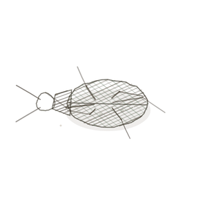
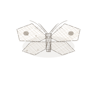
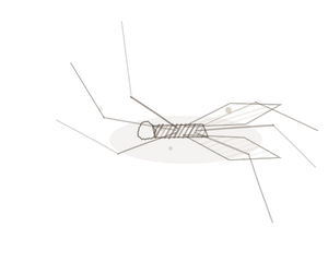
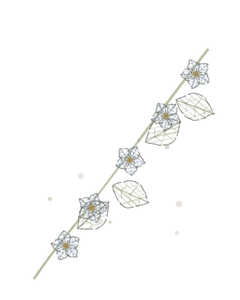
A man o’ war is many individuals fused into one body all pulling the same way. An aspen grove is one organismA living individual made up of cells. branching underground, so a signal in one stem is a signal in all of them. A strangler fig is a slow squeeze you cannot argue with. Those are skeletal, cardiac and smooth muscle, in that order, and you already know all three.
Begin · Pl. I
Striatus multinucleatus var. physalius
the rowing crew · skeletal muscle.
Hop to:
← Epithelial Tissues: A Guided Journey
All plates — The Tissue Field Guide: Epithelium
Frontispiece Ordo Epithelia (main page)
Pl. I · Simple squamous
Pl. II · Simple cuboidal
Pl. III · Simple columnar
Pl. IV · Stratified squamous
Pl. V · Stratified cuboidal
Pl. VI · Stratified columnar
Pl. VII · Transitional
Pl. VIII · Pseudostratified columnar
Epithelial Identification Game
All Modules
- Anatomical Language, Membranes & Homeostasis
- Just Enough Chemistry
- The Cell & Its Transport
- Making Cells & Proteins
- The Integumentary System
- The Skeletal System
- The Muscular System
- Nervous Tissue & the Senses
- The Spinal Cord
- The Brain & the Blood–Brain Barrier
- The Autonomic Nervous System
- The Special Senses
- The Endocrine System
- Blood
- The Heart
- Blood Vessels
- The Digestive System
- The Respiratory System
- The Urinary System
- Fluids, Electrolytes & Acid–Base Balance
- The Reproductive System
- The Immune System
List of terms
- nuclei
- energy
- myofibril
- cells
- dark band
- nucleus
- intercalated discs
- collagen
- organism