Time To Read
Date Last Modified
Pl. II · FAM. LAXIDAE
Laxus adiposus var. mellifer
the vault · adipose tissue
The plate
The specimen, drawn


Habitat & range
Where it lives
Body map

Adipose sits where the body wants either a fuel store, a cushion, or a blanket — and often all three at once.
Range
- The subcutaneous layer, under the whole skinThe body’s largest organ, providing protection and regulation. — the layer you can pinch.
- A packing cushion around each kidney, holding it in place.
- The breast, and the omental apron hanging over the gut.
- Behind each eyeball, and in the palms and soles as shock padding.
- Brown fat between the shoulder blades in newborns, for heat rather than storage.
Collection record
- Add your slide numbers here.
Niche
What it does for a living
Storage, insulation, and padding — and, lately, hormone signalling. An adipocyte is a cell that has committed almost its whole volume to one lipid droplet. It is not inert packing: adipose releases leptinA hormone involved in regulating energy balance and puberty onset. and other signals that tell the brain how much fuel is on hand, and brown fat burns its own stores to make heat instead of ATPThe energy currency of cells used for muscle contraction..
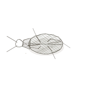
field collected specimen
How to know it on sight
- Empty-looking circles — the fat dissolved out in processing and left the space behind.
- Each cell is a thin rim of cytoplasm around that space, like a signet ring.
- Nuclei squashed flat and pushed against one side, never central.
- Cells packed close with almost no matrix between them; capillariesThe smallest blood vessels where gas, nutrient, and waste exchange occurs between blood and tissues. squeeze through the corners.

Specimen 1

From a recto-anal junction. Find one cell and trace the rim all the way round; the nucleus is the bulge on one side.
Specimen 2

H&E. The clear spaces are where lipid used to be before the solvents took it.
Specimen 3

High power. Look at how little actual cell there is around each space.

People keep calling her by the wrong name
Look-alikes
Two plates. Read the line underneath each one before you decide.
Areolar connective tissue

Areolar has open space too, but it is unbounded gaps between fibres; adipose space is bounded — every gap has its own cell wall round it.
Spongy bone and marrow

Marrow has fat cellsThe basic structural and functional units of life. in it, so the circles look familiar — but marrow also has bony trabeculae and developing blood cells, and adipose has neither.
Examine it yourself
The virtual slide
This is brown fat, not the usual white. Compare it with the slides above: the droplets are many and small rather than one and large, and the nucleus is still roughly central. Ask yourself what burning fat for heat requires that storing it does not.
Virtual slide courtesy of Histology Guide.
YOur missions
Field Notes for Future Missions
Mission 1: find the rim
Mission 2: quick check
Field notes
Why this organism
A honeypot ant replete spends its life hanging from the roof of the nest with its abdomen swollen into a translucent globe of food. It cannot walk, it cannot forage, it cannot do anything else — it has given over its whole body to being a container, and the colony lives off it when the season turns. An adipocyte makes exactly the same bargain: everything that was cell gets pushed to the edges so the cargo can have the middle.
Next · Pl. III
Laxus reticularis var. mycelialis
the invisible net · reticular connective tissue.
Hop to:
← Epithelial Tissues: A Guided Journey
All plates — The Tissue Field Guide: Epithelium
Frontispiece Ordo Epithelia (main page)
Pl. I · Simple squamous
Pl. II · Simple cuboidal
Pl. III · Simple columnar
Pl. IV · Stratified squamous
Pl. V · Stratified cuboidal
Pl. VI · Stratified columnar
Pl. VII · Transitional
Pl. VIII · Pseudostratified columnar
Epithelial Identification Game
All Modules
- Anatomical Language, Membranes & Homeostasis
- Just Enough Chemistry
- The Cell & Its Transport
- Making Cells & Proteins
- The Integumentary System
- The Skeletal System
- The Muscular System
- Nervous Tissue & the Senses
- The Spinal Cord
- The Brain & the Blood–Brain Barrier
- The Autonomic Nervous System
- The Special Senses
- The Endocrine System
- Blood
- The Heart
- Blood Vessels
- The Digestive System
- The Respiratory System
- The Urinary System
- Fluids, Electrolytes & Acid–Base Balance
- The Reproductive System
- The Immune System
List of terms
- cytoplasm
- nucleus
- skin
- leptin
- ATP
- capillaries
- cells