Time To Read
Date Last Modified
Pl. II · FAM. STRIATIDAE
Striatus ramosus var. populinus
the grove · cardiac muscle
The plate
The specimen, drawn
Pl. I
[ plate drawing to come · the aspen grove ]

Habitat & range
Where it lives
Body map
[ body map to come — needs the BioRender export ]
Range
- The myocardium — the muscular wall of all four chambers, thickest by far in the left ventricle.
- Thin sleeves of muscle running a short way up the great veinsBlood vessels that return deoxygenated blood to the heart (except pulmonary veins, which carry oxyge where they enter the atria.
- The conducting cellsThe basic structural and functional units of life. — SA node, AV node, bundle branches, Purkinje fibers. These are modified cardiac muscle, not nerve, which surprises everyone.
- And nowhere else at all. It is the only tissue in the body that lives in a single organ.
Collection record
- Add your slide numbers here.
Niche
What it does for a living
Tireless and automatic. Cardiac muscle contracts somewhere around three billion times in an average life without ever once being told to start, and it is not allowed to fatigue or to fire out of step. The cell is built for exactly that: branched, so force and signal spread sideways as well as end to end; joined to its neighbours by intercalated discs Structures in cardiac muscle that allow electrical connectivity. that are glue and electrical doorway in the same structure; and so packed with mitochondria that it runs aerobically or it does not run. There is no anaerobicprocess that does not use oxygen option and no rest day.
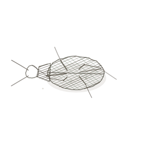
field collected specimen
How to know it on sight
- Striations, as in skeletal muscle — but fainter, and the fibers are far shorter.
- Fibers branch and rejoin, so the tissue reads as a net rather than a bundle of cables.
- One nucleusThe control center of the cell that contains DNA and directs cellular activities. to a cell, round or oval, sitting in the middle of the fiber rather than at its edge.
- Intercalated discs — dark lines crossing a fiber at intervals, marking where one cell stops and the next starts.

Specimen 1

Right heart wall. The fibers fork and rejoin, and the nucleiClusters of neurons in the CNS responsible for processing information. sit in the middle of the fiber, not along its rim.
Specimen 2

Ventricle. Look for dark lines running straight across a fiber — those are intercalated discs, the join between one cell and the next.
Specimen 3

Masson’s trichrome. The muscle takes the red; the blue is the collagenA structural protein in the dermis that provides strength and elasticity. scaffold the fibers are slung in.


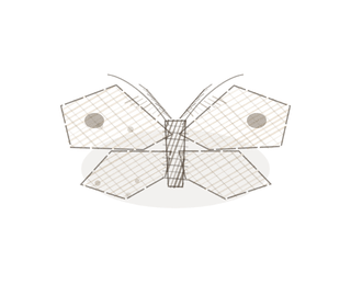
People keep calling her by the wrong name
Look-alikes
Two plates. Read the line underneath each one before you decide.
Skeletal muscle

Tell them apart by the branch. Skeletal fibers run straight and parallel with many nuclei at the rim; cardiac fibers fork, and carry one nucleus in the centre.
Smooth muscle

Tell them apart by the stripes. Both keep one central nucleus, but only cardiac is striated — and smooth muscle never branches.
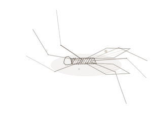
Examine it yourself
The virtual slide
Find a fiber that forks. Then follow it along until you meet a dark line crossing it at right angles: that is an intercalated disc.
Virtual slide courtesy of Histology Guide.
YOur missions
Field Notes for Future Missions
Mission 1: find the discs
Mission 2: quick check
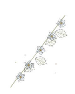
Field notes
Why this organism
Pando is a single quaking aspen in Utah — some forty thousand trunks that look like a forest and are one organismA living individual made up of cells., every stem grafted to its neighbours underground and sharing whatever it has. Cardiac muscle is a grove. The fibers branch and rejoin, and the intercalated discs between them are root grafts: mechanical anchor and open electrical door in one structure, so a signal that starts in one cell does not stay in that cell. That is why a heart beats as a single thing instead of as a bag of cells all twitching on their own schedule.
Next · Pl. III
Laevis fusiformis var. strangulans
the slow squeeze · smooth muscle.
Hop to:
← Epithelial Tissues: A Guided Journey
All plates — The Tissue Field Guide: Epithelium
Frontispiece Ordo Epithelia (main page)
Pl. I · Simple squamous
Pl. II · Simple cuboidal
Pl. III · Simple columnar
Pl. IV · Stratified squamous
Pl. V · Stratified cuboidal
Pl. VI · Stratified columnar
Pl. VII · Transitional
Pl. VIII · Pseudostratified columnar
Epithelial Identification Game
All Modules
- Anatomical Language, Membranes & Homeostasis
- Just Enough Chemistry
- The Cell & Its Transport
- Making Cells & Proteins
- The Integumentary System
- The Skeletal System
- The Muscular System
- Nervous Tissue & the Senses
- The Spinal Cord
- The Brain & the Blood–Brain Barrier
- The Autonomic Nervous System
- The Special Senses
- The Endocrine System
- Blood
- The Heart
- Blood Vessels
- The Digestive System
- The Respiratory System
- The Urinary System
- Fluids, Electrolytes & Acid–Base Balance
- The Reproductive System
- The Immune System
List of terms
- veins
- cells
- intercalated discs
- anaerobic
- nucleus
- nuclei
- collagen
- organism