Time To Read
Date Last Modified
Pl. II · FAM. NEPHRONIDAE
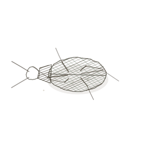
Corpusculum fenestratum var. cyathi
the sieve in a cup · renal corpuscleThe structure in the nephron that consists of the glomerulus and Bowman’s capsule, where filtration
The plate
The specimen, drawn
Pl. II

The fungus as it was drawn, with the corpuscle below it and the filtrationThe process by which fluid moves out of capillaries into surrounding tissues due to hydrostatic pres barrier in the circle.

Habitat & range
Where it lives
Body map

Cortex only. A million of them in each kidney, and never one in the medulla.
Range
- Cortex only, never medulla — about a million per kidney, 200 µm across.
- Two poles: the vascular pole where the arteriolesSmall arteries that regulate blood flow into capillaries through vasoconstriction and vasodilation enter and leave, the urinary pole where the tubule begins.
- Filters roughly 125 mL a minute, which comes to 180 litres a day.
- The only capillary bed in the body with an arteriole at both ends.
Collection record
- Add your slide numbers here.
Niche
What it does for a living
It is a sieve run at high pressureThe force exerted by gases in the respiratory system, affecting airflow and gas exchange., and it does not choose. Three layers — fenestrated endotheliumThe innermost layer of blood vessels, composed of simple squamous epithelial cells, which reduces fr, fused basement membrane, podocyte slit diaphragms — sort by size and by charge, and by nothing else. Anything smaller than about 70 kDa goes through; albuminA plasma protein that helps maintain osmotic pressure and transport substances. is just over the line and is also negative, so it is turned away twice. Net filtration pressure is only about 10 mmHg, which sounds small until you remember it runs every minute of your life. An arteriole at both ends is what lets the kidney hold its filtration rate steady while the blood pressure moves around it.
field collected specimen
How to know it on sight
- A ball of capillariesThe smallest blood vessels where gas, nutrient, and waste exchange occurs between blood and tissues. sitting in a cup, with a clear space all the way around it. Once you have seen the gap you cannot unsee it.
- The parietal layer of Bowman’s capsule is simple squamous — a thin dark line around the outside.
- The visceral layer is podocytesSpecialized cells in the glomerulus that form filtration slits, preventing large molecules from ente wrapped onto the capillaries; at this power they are just the nucleiClusters of neurons in the CNS responsible for processing information. sitting on the tuft.
- At the vascular pole, two arterioles — and the afferent is wider than the efferent. That difference is the pressure.

Specimen 1

Start with the gap. Trace the capsular spaceThe space between the glomerular capsule and the glomerulus where filtrate collects. all the way round before you look at anything else.
Specimen 2

The tuft at higher power. The dark dots on it are podocyte and endothelial nuclei, not separate cellsThe basic structural and functional units of life. sitting on top.
Specimen 3

Fenestrated capillaries in the tuft. You cannot resolve a window at this power — but you can see how much blood is in there.


People keep calling her by the wrong name
Look-alikes
Two plates. One is a thing that is not a corpuscle, and one is a corpuscle that has stopped working.
A cluster of cut tubules

Round profiles packed together can look like a tuft at low power. There is no capsule and no space around them — that is the whole test.
A corpuscle in disease

A normal glomerulusA network of capillaries in the nephron where blood filtration occurs. beside an amyloid deposit — salmon-red on Congo red, apple-green under polarised light. This is the slide that explains Stina’s proteinuria.
Examine it yourself
The virtual slide
Go to a named Renal Corpuscle view and find the vascular pole — the side where two vessels come and go rather than one tubule leaving. Then measure the two arterioles against each other by eye and decide which one is the afferent.
Virtual slide courtesy of Histology Guide.
YOur missions
Field Notes for Future Missions
Mission 1: label the filter
Mission 2: quick check
Field notes
Why this organism
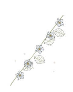
A bird’s nest fungus is a cup with a tangled ball sitting loose inside it and a clear space all the way around — which is the renal corpuscle, structurally, with nothing left out. The fungus earns its place for a second reason: the cup is a splash-cup, and the whole arrangement exists to make waterThe universal solvent essential for life. arriving under pressure do a job. That is the corpuscle’s entire argument.
Next · Pl. III
Tubulus villosus var. callistemonis
the greedy one · proximal convoluted tubuleThe first part of the nephron tubule where most reabsorption of water, ions, and nutrients occurs..
In the wild · photograph to come.
Hop to:
← Epithelial Tissues: A Guided Journey
All plates — The Tissue Field Guide: Epithelium
Frontispiece Ordo Epithelia (main page)
Pl. I · Simple squamous
Pl. II · Simple cuboidal
Pl. III · Simple columnar
Pl. IV · Stratified squamous
Pl. V · Stratified cuboidal
Pl. VI · Stratified columnar
Pl. VII · Transitional
Pl. VIII · Pseudostratified columnar
Epithelial Identification Game
All Modules
- Anatomical Language, Membranes & Homeostasis
- Just Enough Chemistry
- The Cell & Its Transport
- Making Cells & Proteins
- The Integumentary System
- The Skeletal System
- The Muscular System
- Nervous Tissue & the Senses
- The Spinal Cord
- The Brain & the Blood–Brain Barrier
- The Autonomic Nervous System
- The Special Senses
- The Endocrine System
- Blood
- The Heart
- Blood Vessels
- The Digestive System
- The Respiratory System
- The Urinary System
- Fluids, Electrolytes & Acid–Base Balance
- The Reproductive System
- The Immune System
List of terms
- renal corpuscle
- filtration
- arterioles
- pressure
- endothelium
- albumin
- capillaries
- podocytes
- nuclei
- capsular space
- cells
- glomerulus
- water
- proximal convoluted tubule