Time To Read
Date Last Modified
Pl. III · FAM. COLANTIDAE
Colans lymphae var. hydropsychae
the net in the current · lymph node
The plate
The specimen, drawn
Pl. III

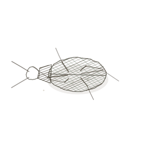
The caddisfly and its net as they were drawn, with a whole lymph node below and the mesh inside one of its sinuses in the circle.

Habitat & range
Where it lives
Body map

Strung along the lymphatic vessels, in clusters, wherever the vessels come together.
Range
- Several hundred of them, from the size of a pinhead to the size of a bean.
- Clustered at the roots of the limbs (armpit, groin), along the neck, and deep in the chest and abdomen beside the great vessels.
- Always in the path of a lymphatic vessel (Pl. II). Lymph passes through at least one node, and usually several, on its way back to the blood.
- The fine net that holds every node together is reticular connective tissue, which has a plate of its own: Ordo Conjunctiva, Pl. III.
Collection record
- Add your slide numbers here.
Niche
What it does for a living
Strain the lymph, and show what is caught to the cells that can respond. A lymph node is the only organ that filters lymph. Afferent vessels pierce the capsule all round its convex side and empty into the subcapsular sinus, a narrow space just beneath. From there lymph seeps inward along the trabeculae and through the medullary sinuses to the hilum, where it leaves by one or two efferent vessels. The sinuses are not open pipes. They are crossed by a mesh of reticular cells and fibres (Ordo Conjunctiva, Pl. III), and macrophages sit in the mesh. The lymph slows to a crawl, and bacteria, debris and stray cells are taken out of it. Around the sinuses the lymphocytes are sorted into zones. The outer cortex holds the follicles, which are B-cell territory; a follicle that has met its antigen swells and develops a pale germinal centre. The deeper cortex, the paracortex, is T-cell territory, and it is where lymphocytes enter the node from the blood. The medulla is cords of cells, many of them plasma cells making antibody, with sinuses between. A node that is working gets bigger, which is why you can feel the ones under your jaw when you have a sore throat.
field collected specimen
How to know it on sight
- An oval or bean-shaped organ, complete, with a capsule of dense connective tissue all the way round it.
- A narrow pale space directly under the capsule: the subcapsular sinus. Nothing else in this volume has one.
- Round follicles in the outer zone, some with a pale centre; a dense deeper zone with no follicles; and a looser middle of dark cords and pale channels.
- The spaces hold lymph. Look in a sinus: pale, with scattered cells, and no red cells to speak of.

Specimen 1

The three zones in order from the capsule inward: cortex with its follicles, paracortex, medulla.
Specimen 2

A small node at low power, lying in fat. The pale channels through its middle are the medullary sinuses.
Specimen 3

Silver stain. The black threads are reticular fibres, the net the whole node hangs on, and the purple cells are caught in it.


People keep calling her by the wrong name
Look-alikes
Two other organs that are a mass of small dark nuclei. Check for follicles, then check the spaces.
Thymus

Thymus — dark outside and pale inside as well, but divided into lobules, with no follicles, no sinuses, and no sinus under the capsule. See Pl. V.
Spleen

Spleen — follicles and a capsule, as in a node. But there is no cortex or medulla, the follicles are scattered throughout, and the field between them is red. See Pl. IV.
Examine it yourself
The virtual slide
At the lowest power, find the capsule and follow it all the way round. Come in one step and find the pale gap just beneath it. Then find a follicle in the cortex, and decide whether it has a germinal centre. Last, go to the middle and look into a sinus at high power for the large, pale cells sitting in the mesh.
Virtual slide courtesy of Histology Guide.
YOur missions
Field Notes for Future Missions
Mission 1: label the node
Mission 2: quick check
Field notes
Why this organism
Turn over a stone in Five Mile Creek in May and the underside is strung with small nets. Each one is the work of a net-spinning caddisfly larva, an inch of green grub with a dark head, which builds itself a shelter of sand grains and then spins a square of silk mesh across the nearest gap where the water runs. Then it stops. It does not go looking for food. It sits at the edge of its net and lets the current bring everything in the stream to it, and whatever is too big to pass through the mesh, it deals with. A lymph node works on the same plan. Lymph is delivered to it; it does not go and fetch any. The sinuses inside are crossed by a mesh, and macrophages sit in that mesh the way the larva sits at its net, taking out of the flow whatever does not belong there. The slower the water, the more the net catches, and lymph in a node barely moves. The mesh itself is reticular tissue, which you have met on Ordo Conjunctiva, Pl. III. One honest difference. The caddisfly eats what it catches, and that is the end of it. In a node the catch is also evidence. Fragments of it are shown to the lymphocytes packed round the sinuses, and if one of them recognises a piece, the follicle it lives in lights up, a germinal centre forms, and the whole node swells. The larva only strains the stream. The node strains it and then raises the alarm.
Next · Pl. IV
Colans sanguinis var. coccinellae
the spotted coat · spleen.
In the wild · photograph to come.
Hop to:
← Epithelial Tissues: A Guided Journey
All plates — The Tissue Field Guide: Epithelium
Frontispiece Ordo Epithelia (main page)
Pl. I · Simple squamous
Pl. II · Simple cuboidal
Pl. III · Simple columnar
Pl. IV · Stratified squamous
Pl. V · Stratified cuboidal
Pl. VI · Stratified columnar
Pl. VII · Transitional
Pl. VIII · Pseudostratified columnar
Epithelial Identification Game
All Modules
- Anatomical Language, Membranes & Homeostasis
- Just Enough Chemistry
- The Cell & Its Transport
- Making Cells & Proteins
- The Integumentary System
- The Skeletal System
- The Muscular System
- Nervous Tissue & the Senses
- The Spinal Cord
- The Brain & the Blood–Brain Barrier
- The Autonomic Nervous System
- The Special Senses
- The Endocrine System
- Blood
- The Heart
- Blood Vessels
- The Digestive System
- The Respiratory System
- The Urinary System
- Fluids, Electrolytes & Acid–Base Balance
- The Reproductive System
- The Immune System