Time To Read
Date Last Modified
Pl. V · FAM. OPERCULIDAE
Operculum stratificatus var. dionaeus
the trapdoor · epiglottisA flap of cartilage that covers the trachea during swallowing to prevent food from entering the airw
The plate
The specimen, drawn
Pl. V

The specimen as it was drawn, with the wall below it and the lining in the circle.

Habitat & range
Where it lives
Body map

Where this one lives.
Range
- At the root of the tongue, above the laryngeal inlet.
- Elastic cartilageA flexible connective tissue found in joints, the ear, nose, and rib cage. Cartilage can be of three, the same tissue as the external ear and the auditory tube.
- Moves about once a minute while you are awake, and on every single swallow.
- The lingual face meets food; the laryngeal face meets air. One organ, two jobs, two linings.
Collection record
- Add your slide numbers here.
Niche
What it does for a living
It is a lid. It folds back over the laryngeal inlet when you swallow and springs forward the rest of the time, and it has to do that roughly once a minute for a lifetime without ever staying bent. Elastic cartilage is the only tissue that will take that; hyaline would fatigue and fail. The epiglottis is the reason the other four plates in this volume stay dry, which is the argument for putting a lid in a volume about airways.
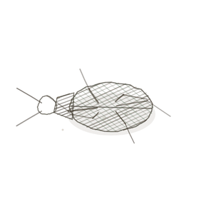
field collected specimen
How to know it on sight
- A core of elastic cartilageA flexible cartilage type found in the ear and epiglottis.: chondrocytes in lacunae, in a matrix stuffed with dark branching elastic fibres — not the clear glassy matrix of the trachea’s hyaline.
- Two different linings on the two faces. Stratified squamous on the lingual side, respiratory pseudostratified ciliated on the laryngeal side.
- A transition between them, somewhere near the tip. Finding it is the best thing to do on this slide.
- Taste buds sometimes on the lingual surface, and sero-mucous glands under the laryngeal side.

Specimen 1

The laryngeal face: pseudostratified, ciliated, the same lining as Pl. I.
Specimen 2

The lingual face: stratified squamous, built to be scraped by food.
Specimen 3

Hyaline cartilage for comparison, in Masson’s trichrome. Note how little the matrix has going on in it.


People keep calling her by the wrong name
Look-alikes
Two plates. Read the line underneath each one before you decide.
Hyaline cartilage

Tell them apart by the matrix, not the cellsThe basic structural and functional units of life.. Hyaline is clear and glassy; elastic is full of dark branching fibres. If the section A cut or slice of the body or an organ for study. is stained for elastin there is no argument left to have.
Hyaline with glands

Tell them apart by where you are as well as what you see. Both organs carry cartilage and glands, but only the epiglottis has two different epithelia on its two faces.
Examine it yourself
The virtual slide
Walk along one face until the lining changes, and stop at the moment it does. That boundary is the whole point of this slide — one organ with food on one side of it and air on the other.
Virtual slide courtesy of Histology Guide.
YOur missions
Field Notes for Future Missions
Mission 1: label the wall
Mission 2: quick check
Field notes
Why this organism
A flytrap is a hinged flap that swings across an opening, springs back to exactly the shape it was, and does it again. Both it and the epiglottis are doors that decide what gets in, and both are built to bend an enormous number of times without fatiguing — which is what elastic cartilage is for. One cross-reference worth making: Ordo Conjunctiva Pl. VIII, The Bouncer, is the fennec fox, and it teaches this same tissue. That plate is about elastic cartilage. This one is about the lid it gets built into.
Back · Frontispiece
Ordo Pneumatica
the air-movers · the five specimens.
In the wild · photograph to come.
Hop to:
← Epithelial Tissues: A Guided Journey
All plates — The Tissue Field Guide: Epithelium
Frontispiece Ordo Epithelia (main page)
Pl. I · Simple squamous
Pl. II · Simple cuboidal
Pl. III · Simple columnar
Pl. IV · Stratified squamous
Pl. V · Stratified cuboidal
Pl. VI · Stratified columnar
Pl. VII · Transitional
Pl. VIII · Pseudostratified columnar
Epithelial Identification Game
All Modules
- Anatomical Language, Membranes & Homeostasis
- Just Enough Chemistry
- The Cell & Its Transport
- Making Cells & Proteins
- The Integumentary System
- The Skeletal System
- The Muscular System
- Nervous Tissue & the Senses
- The Spinal Cord
- The Brain & the Blood–Brain Barrier
- The Autonomic Nervous System
- The Special Senses
- The Endocrine System
- Blood
- The Heart
- Blood Vessels
- The Digestive System
- The Respiratory System
- The Urinary System
- Fluids, Electrolytes & Acid–Base Balance
- The Reproductive System
- The Immune System
List of terms
- epiglottis
- cartilage
- elastic cartilage
- cells
- section