Time To Read
Date Last Modified
Pl. V · FAM. ERUDIENTIDAE
Erudiens lobulatus var. ascidiae
the school that closes · thymus
The plate
The specimen, drawn
Pl. V

The sea squirt and its larva as they were drawn, with the thymus of a child below and a Hassall’s corpuscle in the circle.

Habitat & range
Where it lives
Body map

In the upper chest, behind the breastbone and in front of the great vessels of the heart.
Range
- Two lobes, lying in the mediastinum over the top of the heart.
- Largest relative to the body at birth; largest in absolute terms around puberty.
- After that it is steadily replaced by fat. In an older adult there are islands of thymus in a pad of adipose tissue.
- It receives no lymph. Its cells arrive from the bone marrow (Pl. VI) by the blood, and leave the same way.
Collection record
- Add your slide numbers here.
Niche
What it does for a living
Turn untrained cells from the bone marrow into T lymphocytes, and destroy the ones that turn out wrong. The thymus is a primary lymphoid organ: it makes lymphocytes ready, and does not itself fight anything. Its framework is unusual. It is built of epithelial cells, branched and joined to one another in a sponge, and the lymphocytes are packed into the holes. In the cortex they are packed so tightly that the tissue stains dark. Here each developing T cell is tested twice. Can its receptor recognise the body’s own display molecules at all? If not, it dies. Does it bind the body’s own contents too strongly? Then it would attack you, and it dies as well. Something like ninety-five in every hundred fail and are eaten by macrophages on the spot. The survivors move into the paler medulla and leave by the blood for the lymph nodes and spleen. The medulla also holds the organ’s one unmistakable landmark, the Hassall’s corpuscle: a small whorl of flattened pink epithelial cells, found nowhere else. And because the thymus teaches and does not respond, it has no follicles and no germinal centres. Most of its work is done in childhood, and the organ shrinks once it is.
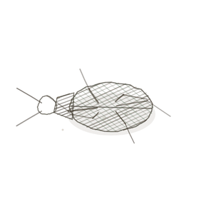
field collected specimen
How to know it on sight
- Lobules: the organ is cut into many-sided blocks by thin septa running in from the capsule.
- In every lobule a dark, crowded rim and a pale core. The pale cores join up from one lobule to the next.
- Hassall’s corpuscles in the pale medulla: round pink whorls, like a cut onion. One of these settles the identification.
- No follicles, no germinal centres and no sinuses. In an adult specimen, fat cells where the lobules used to be.

Specimen 1

The same organ, drawn, so you can see the parts the photograph is too small to show: septa, cortex, medulla, corpuscles.
Specimen 2

Adipose tissue. This is what replaces the thymus with age, so an adult slide is mostly this with islands of lymphoid tissue in it.
Specimen 3

For contrast, a lymph node. One organ with one outer cortex, not a set of lobules, and it has follicles.


People keep calling her by the wrong name
Look-alikes
Two other organs packed with lymphocytes. Count the lobules, then hunt for a follicle.
Lymph node
Lymph node — a dark outer zone and a paler middle, like one giant lobule. But it has round follicles in the cortex and open sinuses in the medulla, and no Hassall’s corpuscles. See Pl. III.
Spleen

Spleen — a capsule and dark lymphoid tissue, but scattered as round islands in a red field, each with an artery in it. No lobules. See Pl. IV.
Examine it yourself
The virtual slide
This slide carries two thymuses, one from a newborn and one from an adult. Start with the newborn: find the lobules, then the dark rim and pale core of one of them, then search the pale core for a round pink whorl. Now move to the adult section and see how much of it is fat.
Virtual slide courtesy of Histology Guide.
YOur missions
Field Notes for Future Missions
Mission 1: label the lobule
Mission 2: quick check
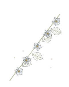
Field notes
Why this organism
A sea squirt starts life as something like a tadpole. It has a head and a tail, a stiffening rod down its back, a nerve cord, an eye spot, and a small brain to steer by, and for a day or so it swims about looking for a good place. Then it glues itself head-first to a rock, and it never moves again. Over the next few days it takes the tadpole apart. The tail goes, the rod goes, most of the nervous system goes, and the material is reused to build what you see on a dock piling: a soft bag with two spouts and a sieve inside. The equipment of its youth did one job, the job is finished, and the animal does not keep it. The thymus is the organ that does this. It is enormous in a baby, hard at work through childhood, and by late middle age most of it has been quietly replaced by fat. Nothing has gone wrong. The T cells it trained are long-lived and are out in the lymph nodes and spleen, and they are still there. One honest difference. The sea squirt does keep a small knot of nerve tissue, and the adult thymus is not quite gone either: it goes on turning out a trickle of new T cells for life. That is why the adult half of your slide still has islands of dark lobule in among the fat.
Next · Pl. VI
Gignens medullaris var. termitis
the nursery in the wall · red bone marrow.
In the wild · photograph to come.
Hop to:
← Epithelial Tissues: A Guided Journey
All plates — The Tissue Field Guide: Epithelium
Frontispiece Ordo Epithelia (main page)
Pl. I · Simple squamous
Pl. II · Simple cuboidal
Pl. III · Simple columnar
Pl. IV · Stratified squamous
Pl. V · Stratified cuboidal
Pl. VI · Stratified columnar
Pl. VII · Transitional
Pl. VIII · Pseudostratified columnar
Epithelial Identification Game
All Modules
- Anatomical Language, Membranes & Homeostasis
- Just Enough Chemistry
- The Cell & Its Transport
- Making Cells & Proteins
- The Integumentary System
- The Skeletal System
- The Muscular System
- Nervous Tissue & the Senses
- The Spinal Cord
- The Brain & the Blood–Brain Barrier
- The Autonomic Nervous System
- The Special Senses
- The Endocrine System
- Blood
- The Heart
- Blood Vessels
- The Digestive System
- The Respiratory System
- The Urinary System
- Fluids, Electrolytes & Acid–Base Balance
- The Reproductive System
- The Immune System