Time To Read
Date Last Modified
Pl. VI · FAM. DENSIDAE
Densus elasticus var. resurgens
the snapback · elastic connective tissue
The plate
The specimen, drawn
Pl. VI

Elastin instead of collagenA structural protein in the dermis that provides strength and elasticity.. Stretch it and it lengthens; let go and it returns to exactly the shape it started in.

Habitat & range
Where it lives
Body map

Elastic tissue goes wherever something has to stretch and come back, over and over, for a lifetime.
Range
- The wall of the aorta and its large branches.
- The ligamenta flava, between the vertebral arches.
- The vocal ligaments.
- The lungs and the bronchial tree.
- The suspensory ligamentA structure that supports the ovary and contains blood vessels. of the penis, and the elastic layers of large airways.
Collection record
- Add your slide numbers here.
Niche
What it does for a living
Storing a push and giving it back. When the heart ejects, the aorta widens and takes some of that volume; when the heart relaxes, the aorta narrows again and keeps the blood moving forward. That is elastic recoil doing a second heartbeat between beats. Elastin is made mostly in youth and barely replaced, which is a large part of why arteriesBlood vessels that carry oxygenated blood away from the heart (except pulmonary arteries, which carr stiffen with age.
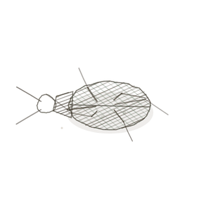
field collected specimen
How to know it on sight
- Dark wavy fibres — black or brown in Verhoeff, refractile in H&E.
- Wavy because they are relaxed; stretch the tissue and they straighten out.
- Fibres branch and rejoin, unlike collagen bundles.
- Often in layers or concentric sheets rather than bundles, especially in vessel walls.

Specimen 1

Verhoeff stain. The dark network is elastin, and it is wavy because nothing is pulling on it at the moment.
Specimen 2

A large blood vessel. The lumenThe inside space of a hollow organ or structure. is at one edge, bounded by an undulating internal elastic lamina A layer of elastic fibers between the tunica intima and tunica media in arteries, allowing for stre.

People keep calling her by the wrong name
Look-alikes
Two plates. Read the line underneath each one before you decide.
Dense regular connective tissue

Collagen bundles are thick, straight and pink. Elastic fibres are thinner, wavy, and dark in an elastic stain.
Dense irregular connective tissue

Dense irregular is bundles with no direction; elastic tissue usually keeps a direction, it is just a wavy one.
Examine it yourself
The virtual slide
Mesentery spread thin enough to show individual fibres. Look for the thin dark lines that branch and rejoin — those are elastic. The thick pale bundles that never branch are collagen.
Virtual slide courtesy of Histology Guide.
YOur missions
Field Notes for Future Missions
Mission 1: wavy or straight
Mission 2: quick check
Field notes
Why this organism
A resurrection plant dries to a brown fist and sits that way for years, and then an hour of rain opens it flat and green again. Nothing was rebuilt; the shape was stored in the thing the whole time. Elastin is that trick done seventy times a minute for eighty years. Your aorta has been doing it since before you were born and has never once had to be told what shape to go back to.
Next · Pl. VII
Sustinens hyalinus var. tremellus
the glider · hyaline cartilageThe most abundant cartilage type, found in joints, ribs, and the nose..
Hop to:
← Epithelial Tissues: A Guided Journey
All plates — The Tissue Field Guide: Epithelium
Frontispiece Ordo Epithelia (main page)
Pl. I · Simple squamous
Pl. II · Simple cuboidal
Pl. III · Simple columnar
Pl. IV · Stratified squamous
Pl. V · Stratified cuboidal
Pl. VI · Stratified columnar
Pl. VII · Transitional
Pl. VIII · Pseudostratified columnar
Epithelial Identification Game
All Modules
- Anatomical Language, Membranes & Homeostasis
- Just Enough Chemistry
- The Cell & Its Transport
- Making Cells & Proteins
- The Integumentary System
- The Skeletal System
- The Muscular System
- Nervous Tissue & the Senses
- The Spinal Cord
- The Brain & the Blood–Brain Barrier
- The Autonomic Nervous System
- The Special Senses
- The Endocrine System
- Blood
- The Heart
- Blood Vessels
- The Digestive System
- The Respiratory System
- The Urinary System
- Fluids, Electrolytes & Acid–Base Balance
- The Reproductive System
- The Immune System
List of terms
- collagen
- suspensory ligament
- arteries
- lumen
- internal elastic lamina
- hyaline cartilage