Time To Read
Date Last Modified
Pl. X · FAM. RECIPIENTIDAE
Recipiens cyclicus
The Sap Run · uterusThe muscular organ where a fertilized egg implants and develops.
The plate
The specimen, drawn
Pl. X

The tree in sap season, with the endometriumThe inner lining of the uterus that thickens during the menstrual cycle to support a potential pregn drawn twice below it at one scale, and a secretory gland read at ×100 in the circle.

Habitat & range
Where it lives

Range
- Central in the pelvic cavityThe body cavity that houses the reproductive organs and bladder., anteverted over the bladder, with the rectum behind it.
- About 7 cm long in a non-pregnant adult; fundusThe dome-shaped upper part of the uterus., body, and the cervix below.
- Three layers: perimetriumThe outermost layer of the uterus. outside, the massive myometriumThe thick muscular layer of the uterus, and the endometrium lining the cavity.
- The endometrium is rebuilt and shed roughly every 28 days, about 400 times in a lifetime.
Collection record
- Add your slide numbers here.
Niche
What it does for a living
Build a surface good enough to implant on, once a month, on spec — and then take it down again without destroying the ground it was built on.
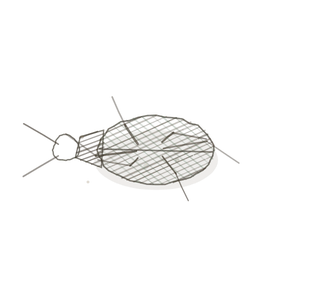
field collected specimen
How to know it on sight
- Two endometrial layers: a functional layerThe superficial layer of the endometrium that is shed during menstruation. that cycles, and a permanent basal layerThe deeper layer of the endometrium that remains intact during menstruation and regenerates the func underneath that regrows it.
- Proliferative phase: a thin functional layer with straight, narrow, sparse tubular glands. Oestrogen did this.
- Secretory phase: a deepAway from the surface of the body. functional layer with coiled, saw-toothed glands full of glycogenA storage form of glucose found in animals.. Progesterone did this.
- Spiral arteriesBlood vessels that carry oxygenated blood away from the heart (except pulmonary arteries, which carr elongate and coil deep into the functional layer in the secretory phaseThe phase of the menstrual cycle when the endometrium is prepared for implantation., and are hard to find before it.

Specimen 1 · proliferative
Straight, narrow glands in a relatively thin functional layer. Oestrogen from the growing follicle is running the show.
Specimen 2 · secretory

A thickened functional layer with elongated, coiled glands and prominent spiral arteriesBlood vessels supplying the endometrium, shedding during menstruation.. There is a corpus luteum behind this.
Specimen 3 · menstrual

The functional layer breaking down, with the glands elongating and the lining beginning to come away.


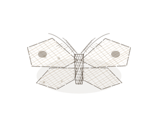
People keep calling her by the wrong name
Look-alikes
The pair that matters most in this volume is not two organs but one organ twice. Learn to call the phase and you can say what the ovary was doing the week the biopsy was taken. The second plate is the doorway below, which never cycles like this.
Proliferative vs secretory
Coiled, saw-toothed, secreting glands and deep spiral arteries mean progesteroneA hormone that supports pregnancy and regulates the menstrual cycle., which means a working corpus luteum.
Cervix

Simple columnar again, but secreting mucus, with no functional layer to shed and no spiral arteries to lose.
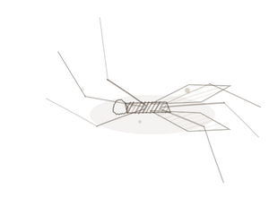
Examine it yourself
The virtual slide
Centre the endometrium only and go to about ×1. Decide the phase from the glands before you look at anything else, then check your answer against the spiral arteries near the top of the functional layer.
Virtual slide courtesy of Histology Guide.
YOur missions
Field Notes for Future Missions
Mission 1: name the phase
Mission 2: quick check

Field notes
Why this organism
A sugar maple spends the whole winter looking like nothing is happening, and then for about six weeks it runs sweet sap under pressureThe force exerted by gases in the respiratory system, affecting airflow and gas exchange. — and only then, and only if the temperature swings the right way. Tap it in August and you get nothing. The endometrium has the same two faces: the proliferative phase The phase when the endometrium rebuilds after menstruation. is a tree building, the secretory phase is the sap run, and a pathologist can name the week from a biopsy exactly the way a sugarmaker can name the season from a bucket.
Next · Pl. XI
Recipiens transiens
The Borderline · cervix.
In the wild · photograph to come.
Hop to:
← Epithelial Tissues: A Guided Journey
All plates — The Tissue Field Guide: Epithelium
Frontispiece Ordo Epithelia (main page)
Pl. I · Simple squamous
Pl. II · Simple cuboidal
Pl. III · Simple columnar
Pl. IV · Stratified squamous
Pl. V · Stratified cuboidal
Pl. VI · Stratified columnar
Pl. VII · Transitional
Pl. VIII · Pseudostratified columnar
Epithelial Identification Game
All Modules
- Anatomical Language, Membranes & Homeostasis
- Just Enough Chemistry
- The Cell & Its Transport
- Making Cells & Proteins
- The Integumentary System
- The Skeletal System
- The Muscular System
- Nervous Tissue & the Senses
- The Spinal Cord
- The Brain & the Blood–Brain Barrier
- The Autonomic Nervous System
- The Special Senses
- The Endocrine System
- Blood
- The Heart
- Blood Vessels
- The Digestive System
- The Respiratory System
- The Urinary System
- Fluids, Electrolytes & Acid–Base Balance
- The Reproductive System
- The Immune System
List of terms
- uterus
- endometrium
- bladder
- rectum
- pelvic cavity
- fundus
- perimetrium
- myometrium
- functional layer
- basal layer
- deep
- glycogen
- arteries
- secretory phase
- spiral arteries
- progesterone
- pressure
- proliferative phase