Time To Read
Date Last Modified
Frontispiece · ORD. CUSTODIA
Ordo Custodia
the sentries · the lymphatic system
The plates
The seven specimens
Seven plates: two kinds of vessel that drain the tissues, and five places where lymphocytes are made, trained or stationed. Two relatives live in other volumes. The tonsils and Peyer’s patches, the appendix’s partners in the wall of the gut, are on Ordo Alimentaria, Pl. XI. The fine net that holds a lymph node and the spleen together is reticular connective tissue, on Ordo Conjunctiva, Pl. III.

Pl. II
Hauriens moniliformis var. hormosirae
the string of beads · collecting lymphatic vessel


The ground plan
Cells that wait, and the fluid that is brought to them
The lymphatic system is two things working together. One is plumbing: thin vessels that collect the fluid blood capillaries leave behind in the tissues and carry it back to the veins. The other is a population of lymphocytes, small round cells that are nearly all nucleus, parked at fixed stations along the way and waiting for something to recognise. Where they gather thickly they form a follicle, and the follicle is the unit this whole volume is built from.
Fig. 1 · the follicle, at rest and at work

A dark ball is resting. A dark ring round a pale centre has met something.
The five words
- Lymph — tissue fluid, once it is inside a lymphatic vessel. Pale, watery, and carrying a few lymphocytes.
- Lymphocyte — a small round cell, nearly all nucleus. Packed together they make a tissue look dark purple.
- Follicle (or nodule) — a round, crowded ball of lymphocytes, mostly B cells.
- Germinal centre — the pale middle a follicle develops when its B cells have met an antigen and begun dividing.
- Capsule — a coat of dense connective tissue round an organ. Some of these have one and some pointedly do not.
So before anything else, find the small dark nuclei, and ask whether they are gathered into follicles.

First question
Is it a tube, or is it a mass?
Two of the seven are vessels. They are easy to walk past, because a lymphatic looks like an empty vein.
Fig. 2 · three thin tubes

Look at what is inside before you look at the wall.
- A thin-walled channel that begins at a closed end, with overlapping cells and nothing red inside — a lymphatic capillary: Pl. I.
- A thin-walled, beaded vessel with a valve at every pinch and pale contents — a collecting lymphatic: Pl. II.
- Red cells inside — a blood vessel, however thin its wall. It belongs to Ordo Vasa, not here.
- And if it is not a tube at all but a mass of small dark nuclei, go on to the second question.
This one question names two of the seven, and it is settled by the contents: pale and nearly empty is lymph, red is blood.
Second question
A mass of lymphocytes: which one?
Three checks, in any order: is there a capsule, are there follicles, and what is in the spaces between?
Fig. 3 · four masses of lymphocytes, at low power

Capsule, follicles, spaces. Three answers name each one.
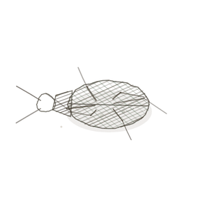
- A capsule and follicles. Follicles at the rim with pale lymph-filled sinuses inside — a lymph node: Pl. III. Follicles round small arteries in a red, blood-filled field — the spleen: Pl. IV.
- A capsule and lobules, each dark at the rim and pale at the core, and no follicles at all — the thymus: Pl. V.
- No capsule. Follicles sitting directly under an epithelium, in the wall of a tube — MALT. In a complete ring round a small lumen it is the appendix: Pl. VII. For tonsils and Peyer’s patches see Ordo Alimentaria, Pl. XI. And fat cells with every sort of cell mixed between them, inside bone, is red marrow: Pl. VI.
Notice what the follicle tells you. Present, and where: node, spleen, MALT. Absent: thymus, where cells are trained and nothing is fought.

The naming key
How to read a name
Genus is the job the specimen does. Species is the mark you find it by, or what it works on. The var. is the organism on the plate.
First word · the job
- Hauriens — draining; drawing fluid off
- Colans — straining; filtering what flows through
- Erudiens — schooling
- Gignens — begetting; making new cells
- Vigilans — standing guard
Second word · the mark, or the material
- caecus, moniliformis — blind-ended; like a string of beads
- lymphae, sanguinis — of lymph; of blood
- lobulatus, medullaris — in lobules; of the marrow
- appendicis — of the appendix
Worked both ways
- Colans sanguinis → the one that strains blood → the spleen.
- Hauriens caecus → a drain with a blind end → a lymphatic capillary.
- You saw lobules, each dark outside and pale inside, and a pink whorl in one pale core → Erudiens lobulatus → Pl. V.
Two that trip people up
The look-alikes
Two mistakes account for most of the lost points in this unit. In both, the overall colour is the same on each side, and something small decides it.
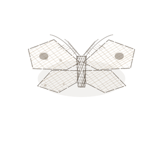
Fig. 4 · two pairs that trip people up

A corpuscle or a follicle settles the first. The contents settle the second.
Thymus, or lymph node?
The trap: both are dark at the edge and paler in the middle.
A lymph node is one organ with one cortex and one medulla. The thymus is many lobules, and every lobule has its own dark rim and pale core. A node has round follicles in its cortex and open sinuses in its medulla. The thymus has neither.
Look in the pale part. A round pink whorl, a Hassall’s corpuscle: thymus. Open channels with cells drifting in them: lymph node.
A lymphatic vessel, or a vein?
The trap: both are thin-walled, irregular, and have valves.
A vein carries blood, so its lumen holds red cells, and its wall, thin as it is, has a visible layer of muscle. A lymphatic of the same size has a wall you can barely see, far more valves, and pale contents with a few lymphocytes.
Look inside first. Red cells: vein. Nothing red, and a bead-and-pinch outline: lymphatic.
Keying out a specimen
Three questions, in this order
This is the procedure. Work down it, and do not skip to the one you think you recognise.
- A tube, or a mass? A thin-walled tube with nothing red inside is a lymphatic. Closed at one end with overlapping cells → capillary. Beaded, with valves → collecting vessel.
- A mass of small dark nuclei: is there a capsule, and are there follicles? Capsule and follicles → lymph node or spleen; go to step 3. Capsule, lobules, no follicles → thymus. No capsule, follicles under an epithelium → MALT; a full ring round a small lumen is the appendix.
- What is in the spaces? Pale lymph, in a sinus under the capsule and through the middle → lymph node. Red cells everywhere, with the follicles as dark islands round small arteries → spleen.
Three overrides. Fat cells with a crowd of mixed cells between them, inside bone, is red marrow, whatever else you think you see. A Hassall’s corpuscle means thymus even when most of the slide is fat. And red cells in the lumen mean it is a blood vessel, however thin the wall.
YOur missions
Field Notes for Future Missions
Before you go to the plates, try the ground plan on a field you have not met yet.
Mission 1 · the follicle, at rest and at work
Mission 2 · key it out
Field notes
Why seven organisms
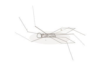
A bladderwort, a seaweed, a caddisfly, a ladybird, a sea squirt, a termite mound and an okra pod have nothing to do with immunity. That is the point. Each one already shows, out in the world, the one structural idea its specimen turns on: a door that only opens inward, a chain of beads, a net set across a current, dark islands in a bright field, a job finished in youth and then dismantled, many kinds from one source inside a hard wall, and a ring of round bodies in the wall of a dead end.
This system is easy to treat as a list of organs that all look like purple dots. They are not alike. Each one is in a different place for a different reason, and on a slide each gives itself away by where its follicles are, by whether it has any, and by what fills the spaces between.
Begin · Pl. I
Hauriens caecus var. utriculariae
the trapdoor · lymphatic capillary.
Hop to:
← Epithelial Tissues: A Guided Journey
All plates — The Tissue Field Guide: Epithelium
Frontispiece Ordo Epithelia (main page)
Pl. I · Simple squamous
Pl. II · Simple cuboidal
Pl. III · Simple columnar
Pl. IV · Stratified squamous
Pl. V · Stratified cuboidal
Pl. VI · Stratified columnar
Pl. VII · Transitional
Pl. VIII · Pseudostratified columnar
Epithelial Identification Game
All Modules
- Anatomical Language, Membranes & Homeostasis
- Just Enough Chemistry
- The Cell & Its Transport
- Making Cells & Proteins
- The Integumentary System
- The Skeletal System
- The Muscular System
- Nervous Tissue & the Senses
- The Spinal Cord
- The Brain & the Blood–Brain Barrier
- The Autonomic Nervous System
- The Special Senses
- The Endocrine System
- Blood
- The Heart
- Blood Vessels
- The Digestive System
- The Respiratory System
- The Urinary System
- Fluids, Electrolytes & Acid–Base Balance
- The Reproductive System
- The Immune System