Time To Read
Date Last Modified
Frontispiece · ORD. FILTRANTIA
Ordo Filtrantia
the filters · the urinary systemThe organ system that removes waste and maintains fluid balance.
The plates
The eight specimens
Four families, and the split between them is the lesson. One plate for the organ, four for the nephronThe functional unit of the kidney that filters blood and produces urine. that actually makes the urineThe liquid waste excreted by the kidneys., one for the collecting system that is not part of the nephron at all, and two for the road out, where nothing is changed any more.
Pl. I
Ren zonatus var. actinidius
the sorting house · kidney

Open Pl. I →
Pl. II
Corpusculum fenestratum var. cyathi
the sieve in a cup · renal corpuscleThe structure in the nephron that consists of the glomerulus and Bowman’s capsule, where filtration

Open Pl. II →
Pl. III
Tubulus villosus var. callistemonis
the greedy one · proximal convoluted tubuleThe first part of the nephron tubule where most reabsorption of water, ions, and nutrients occurs.

Open Pl. III →
Pl. IV
Tubulus tenuis var. ardeae
the long game · nephron loop, thin limb

Open Pl. IV →
Pl. V
Tubulus densus var. mimosae
the sensor · distal tubule and macula densaA group of specialized cells in the distal convoluted tubule that detect sodium concentration and he

Open Pl. V →
Pl. VI
Ductus discretus var. sphagni
the gatekeeper · collecting ductA duct in the nephron that collects urine from multiple nephrons and adjusts water reabsorption.

Open Pl. VI →
Pl. VII
Via stellata var. carambolae
the star · ureterThe tube that carries urine from the kidney to the bladder.

Open Pl. VII →
Pl. VIII
Via trilaminata var. octopodis
the squeeze · bladderA muscular organ that stores urine before excretion.

Open Pl. VIII →
The ground plan
One tube, twice as long as it looks
Every nephron is a single unbranched tube with a filter bolted to the front of it, and almost all of its length is spent taking back what the filter threw away. About 180 litres of plasmaThe liquid component of blood. are filtered a day and about 1.5 litres leave as urine, so roughly 99 % of what crosses the filter is reclaimed further down. Filtering happens only in the cortex. Concentrating happens only in the medulla. Nearly everything else on this page follows from those two facts.
Fig. 1 · one nephron, end to end

Filter first, then three tubules that each take back something different — and the collecting duct in slate blue, because it belongs to a different family.
The five words
- Cortex — the grainy outer zone. Every renal corpuscle in the kidney sits here, which is why filtrationThe process by which fluid moves out of capillaries into surrounding tissues due to hydrostatic pres happens only here.
- Medulla — the striped inner zone, arranged in pyramids. Everything in it is cut lengthwise — loops, collecting ducts and straight vessels, all running the same way.
- Nephron — one corpuscle and its tubules, from the filter to the point where it joins a collecting duct. A million per kidney, and the whole of urine production happens inside one.
- Collecting system — the ducts the nephrons drain into. Not part of the nephron: different embryonic origin, different job, and the only place a hormone finishes the work.
- Filtrate — what crosses the filter. It is not urine yet, and it will not be until the last duct has had its say.
Decide whether you are in cortex or medulla before anything else. It settles half the question on its own.

First question
Where are you?
Before you look at a single cell, work out which zone of the kidney you are in — or whether you have left the kidney altogether.
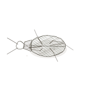
Fig. 2 · where are you

Three fields. Corpuscles, then everything cut lengthwise, then a wall thick enough to be a road.
- Cortex — round corpuscles scattered among tubules cut every which way. One corpuscle anywhere in the field is enough to settle it.
- Medulla — no corpuscles at all, and everything running parallel because it is all cut lengthwise.
- Outside the kidney — a lumenThe inside space of a hollow organ or structure. with a thick muscular wall around it and domed cellsThe basic structural and functional units of life. on the surface. That is the road out, not the kidney.
Corpuscles are the test. Their absence is not proof on its own, because a badly cut cortical field can miss them — so ask whether the tubules are running parallel as well.
Second question
What is the lining doing?
Filtration is indiscriminate: it throws out everything small enough to go. Every tubule after it is choosy, and the lining tells you how choosy.
Fig. 3 · what the lining is doing

Four linings, from taking back nearly everything to taking back nothing at all.
- Brush-bordered — fuzz filling the lumen so you cannot find its edge. Taking back nearly everything: the proximalCloser to the point of attachment or origin. convoluted tubule.
- Bare cuboidal — cube-shaped cells, a clean open lumen, nothing on the free surface. Taking back only sodium(Na⁺): Major ECF cation; important for fluid balance, nerve function., and only on orders: the distal convoluted tubuleThe part of the nephron where final adjustments to ion and water balance occur under hormonal contro.
- Sharp-bordered — pale boxy cells with knife-edged joins between them. Taking back waterThe universal solvent essential for life. only when a hormone says so: the collecting duct.
- Urothelium — several rows of cells with big domed ones on top. Taking back nothing, ever — which is exactly why it is waterproof: ureter and bladder.
A fuzzy lumen beats everything else in the kidney. If you cannot find the edge of the lumen, you have a proximal tubule and nothing more needs deciding.

The naming key
How to read a name
Two words. The first says where you are in the path; the second says what the lining is doing there.
First word · where you are
- Ren — the whole organ
- Corpusculum — the filter itself
- Tubulus — a tubule of the nephron
- Ductus — the collecting system, which is not the nephron
- Via — the road out
Second word · what the lining is doing
- zonatus — zoned — cortex, medulla, pelvis
- fenestratum — windowed — holes straight through
- villosus — shaggy — a brush borderA dense layer of microvilli on the surface of certain cells, increasing surface area for absorption.
- tenuis — one cell thin, and nothing on it
- densus — crowded — cells packed close and sensing
- discretus — sharp — knife-edged borders
- stellata — folded to a star
- trilaminata — three coats of muscle
Worked both ways
- Tubulus villosus → a nephron tubule with a brush border → the proximal convoluted tubule.
- Via stellata → the road out, with a star-shaped lumen → the ureter.
- You saw pale boxy cells with knife-edged borders running through the medulla → Ductus discretus → a collecting duct.
Three that trip people up
The look-alikes
Three pairs account for most of the mistakes in this volume. Each one has a single reliable test, and not one of those tests is size.
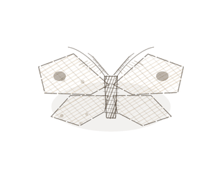
Fig. 4 · three that trip people up

Three pairs, stacked. Lumen, then contents, then wall.
A proximal tubule, or a distal one?
The trap: both are cuboidal, and both sit in the cortex.
They are neighbours in the same field and much the same shape, so position will not separate them. The proximal tubule carries a brush border that fills its lumen with fuzz and makes the edge impossible to find. The distal tubule has a clean open lumen you could draw a line around. The proximal one is also the wider of the two, with fewer nucleiClusters of neurons in the CNS responsible for processing information. in a cross-section, because its cells are bigger.
Look for the edge of the lumen. If you can find it, it is distal.
A thin limb, or a vas rectum?
The trap: both are thin-walled tubes running lengthwise through the medulla.
Both are one flat cell thick and both run with the pyramids, so neither shape nor direction is any help. The vas rectumThe final section of the large intestine, where feces are stored before elimination. is a blood vessel, so there are red blood cells sitting in it. The thin limb is a tubule, so it is empty, or holds nothing but pale filtrateThe fluid that is filtered from the blood into the nephron and will eventually become urine..
Check the contents, not the wall. Red cells inside means a vessel.
A ureter, or a bladder?
The trap: both are lined with urothelium.
The lining is identical in the two, so it cannot separate them. The ureter has a lumen folded into a star, and nothing else in the body has that outline; its wall carries two coats of muscle. The bladder has a wide, shallow lumen and three coats that cross one another, which is what lets it squeeze itself empty from every direction at once.
A star means ureter. Count the muscle coats to confirm it.
Keying out a specimen
Four questions, in this order
This is the procedure. It works on a urinary slide you have never seen before and have not been told the name of, which is the only test that matters.
- Are you inside the kidney at all? — A thick muscular wall around the lumen, with domed cells on the surface, means you have left it — go to Pl. VII or Pl. VIII.
- Cortex or medulla? — One corpuscle anywhere in the field means cortex. No corpuscles, and everything cut lengthwise, means medulla.
- Can you see the lumen clearly? — Full of fuzz, with no findable edge, means a proximal convoluted tubule. Clean and open means a distal tubule or a collecting duct.
- Are the cell borders sharp? — Knife-edged, boxy and pale means a collecting duct, wherever in the kidney you happen to be.
Three overrides. A fuzzy lumen beats everything — a brush border is the single most reliable mark in the kidney. Sharp borders beat location, because collecting ducts run through cortex and medulla alike and look the same in both. Domed surface cells outside the kidney mean urothelium — and then the lumen decides which, because a star is only ever a ureter.
YOur missions
Field Notes for Future Missions
Before you go to the plates, try the key on the whole path at once.
Mission 1 · follow the filtrate down
Mission 2 · key it out
Field notes
Why eight organisms
A kiwifruit, a bird’s nest fungus, a bottlebrush flower, a great blue heron, a sensitive plant, a cushion of sphagnum moss, a starfruit and an octopus have nothing to do with one another and nothing to do with kidneys. That is rather the point of them.
A list of eight structures is something you memorise and then lose. Eight organisms that each carry one structural idea — drainage running outward through productive tissue, a sieve sitting in a cup, a surface multiplied into fuzz, a long thin counterflow, a patch that senses and reports, a gate that opens only when told, a star-shaped lumen, three crossing coats of muscle — stay put, because you can picture them. The name is a handle for the picture, not the other way round.
Begin · Pl. I
Ren zonatus var. actinidius
the sorting house · kidney.
Hop to:
← Epithelial Tissues: A Guided Journey
All plates — The Tissue Field Guide: Epithelium
Frontispiece Ordo Epithelia (main page)
Pl. I · Simple squamous
Pl. II · Simple cuboidal
Pl. III · Simple columnar
Pl. IV · Stratified squamous
Pl. V · Stratified cuboidal
Pl. VI · Stratified columnar
Pl. VII · Transitional
Pl. VIII · Pseudostratified columnar
Epithelial Identification Game
All Modules
- Anatomical Language, Membranes & Homeostasis
- Just Enough Chemistry
- The Cell & Its Transport
- Making Cells & Proteins
- The Integumentary System
- The Skeletal System
- The Muscular System
- Nervous Tissue & the Senses
- The Spinal Cord
- The Brain & the Blood–Brain Barrier
- The Autonomic Nervous System
- The Special Senses
- The Endocrine System
- Blood
- The Heart
- Blood Vessels
- The Digestive System
- The Respiratory System
- The Urinary System
- Fluids, Electrolytes & Acid–Base Balance
- The Reproductive System
- The Immune System
List of terms
- urinary system
- nephron
- urine
- renal corpuscle
- proximal convoluted tubule
- macula densa
- collecting duct
- ureter
- bladder
- plasma
- filtration
- lumen
- cells
- proximal
- sodium
- distal convoluted tubule
- water
- brush border
- nuclei
- rectum
- filtrate