Time To Read
Date Last Modified
Frontispiece · ORD. CONJUNCTIVA
Ordo Conjunctiva
the in-betweeners · connective tissue
The plates
The twelve specimens
Twelve specimens, and almost all of them are told apart by proportion rather than by any single feature. Every one of these tissues is made of the same three things. What changes is how much of each, and which way the fibres run.

Pl. III
Laxus reticularis var. mycelialis
the invisible net · reticular connective tissue


Pl. VII
Sustinens hyalinus var. tremellus
the glider · hyaline cartilageThe most abundant cartilage type, found in joints, ribs, and the nose.

Pl. VIII
Sustinens elasticus var. auritus
the bouncer · elastic cartilageA flexible cartilage type found in the ear and epiglottis.

Pl. IX
Sustinens fibrosus var. nuciferus
the shock absorber · fibrocartilageA tough, shock-absorbing cartilage found in intervertebral discs and menisci.

Pl. X
Sustinens osseus var. corallinus
captain concrete · compact boneDense, strong bone tissue forming the outer layer of bones.

Pl. XI
Sustinens osseus var. trabecularis
the lattice · spongy boneA porous bone tissue found inside bones, providing lightweight support.

Pl. XII
Sustinens transiens var. bambusinus
the shapeshifter · the epiphyseal plateA growth plate in developing bones made of cartilage.

The ground plan
Three ingredients, and the proportions decide
Every connective tissue in this volume is cellsThe basic structural and functional units of life., fibres, and ground substance — and that is the whole list. Epithelium is nearly all cells; connective tissue is nearly all matrix, and the cells are scattered through it like currants. So the questions that identify one of these tissues are not really about the cells at all. They are about how much matrix there is, what is in it, and whether it has a direction.
Fig. 1 · cells, fibres, and the space between

Areolar tissue, the plainest case. A few cells, fibres of all three kinds, and a great deal of space that is not empty but full of ground substance.
The words you need
- Ground substance — the gel the rest of it sits in. Mostly waterThe universal solvent essential for life., held by large sugar-protein moleculesGroups of atoms bonded together.. You cannot see it; you see the space it occupies.
- Collagen — thick, strong, pink in H&E, and the commonest protein in your body. Enormously strong in tension and useless in compression.
- Elastic fibres — thinner, branching, and able to stretch and recoil. They need their own stain to show up properly.
- Reticular fibres — fine branching type III collagenA structural protein in the dermis that provides strength and elasticity. that forms nets rather than bundles. Shown by silver, not by H&E.
- Fibroblast — the cell that makes all of the above. In cartilageA flexible connective tissue found in joints, the ear, nose, and rib cage. Cartilage can be of three it is called a chondrocyteMature cartilage cells housed in lacunae., in bone an osteocyteMature bone cells that maintain bone tissue., and it sits in a little hole called a lacunaSmall spaces in bone tissue where osteocytes reside..
Decide what the matrix is made of before you try to name the tissue. Almost every mistake in this volume comes from naming it first.

First question
Loose, dense, or supporting?
This is the first question and it splits the twelve into three families. Do not look at the cells. Look at how much of the field is fibre and how much is space, and whether the matrix is something you could put your finger through.
Fig. 2 · how much of it is fibre

A dense tissue. Compare the amount of open space here with Fig. 1 — that difference is the whole of the loose-versus-dense distinction.
- More space than fibre → loose, FAM. LAXIDAE. Areolar, adipose, reticular.
- More fibre than space → dense, FAM. DENSIDAE. Dense regular, dense irregular, elastic.
- A solid matrix you could not push a finger through → supporting, FAM. SUSTINENTIDAE. The cartilages, and bone.
Loose and dense are both connective tissue proper, and both have cells you would recognise as fibroblasts. Supporting tissues put their cells in lacunae and change the name.
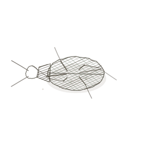
Second question
Which supporting tissue?
If you have decided the matrix is solid, there are only four answers left, and the first question is whether it is mineralised. Bone is hard because of calcium; cartilage is firm because of bound water.
Fig. 3 · matrix, and what is in it
Compact bone. Rings around a central canalA hollow canal in an osteon containing blood vessels and nerves., with cells trapped in lacunae between them — the structure you only need when nothing can diffuse through the matrix.
- Mineralised, with concentric rings around a canal → compact bone.
- Mineralised, as an open lattice of struts → spongy bone.
- Not mineralised, matrix featureless → hyaline cartilage.
- Not mineralised, matrix full of a dark fibre mesh → elastic cartilage.
- Not mineralised, thick visible bundles with cells in rows → fibrocartilage.
And one case that is none of these: a band with cartilage at one end and bone at the other, in five zones. That is the epiphyseal plate, and it is a place rather than a tissue.

The naming key
How to read a name
Put the two answers together — how much matrix first, then what the matrix is doing — and the name writes itself. The nicknames in this volume are the ones you already know from the In-Betweeners; the binomials are new.
First word · how much matrix, and how solid
- Laxus — loose. More space than fibre.
- Densus — dense. More fibre than space.
- Sustinens — supporting. A solid matrix, and the cells sit in lacunae.
Second word · the pattern
- vagus — wandering. Fibres every way, and mostly space (areolar).
- adiposus — cells that have given their whole volume to one fat droplet.
- reticularis — a fine branching net rather than bundles.
- rectus — bundles all running one way.
- confusus — bundles running every way at once.
- elasticus — elastin. Stretches, and returns.
- hyalinus — glassy. No fibres visible at all.
- fibrosus — thick bundles, with cartilage cells in rows between them.
- osseus — mineralised. Rings for compact, struts for spongy.
- transiens — in transition. Cartilage at one end, bone at the other.
Worked both ways
- Sustinens hyalinus → supporting, glassy matrix → hyaline cartilage.
- Densus confusus → dense, no grain → dense irregular, which is your dermisThe thick inner layer of the skin that contains blood vessels, nerves, and connective tissue..
- You saw empty circles packed edge to edge with flattened nucleiClusters of neurons in the CNS responsible for processing information. → Laxus adiposus → adipose.
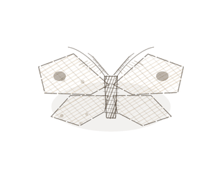
Two that trip people up
The look-alikes
Neither of these breaks the key. They catch people out because the pairs look identical until you ask the one question that separates them — so ask it first, before you have talked yourself into an answer.
Fig. 4 · the pair that settles it

Dense regular. Every bundle in this field shares one direction — which is exactly what dense irregular never does.
Dense regular mistaken for dense irregular
Both are packed solid with thick pink collagen bundles and very little else.
The density is the same, the stain is the same, and the cells are the same. The only difference is whether the bundles agree about direction.
Find two bundles crossing. If you can, it is irregular. If every bundle in the field runs the same way, it is regular.
Hyaline cartilage mistaken for fibrocartilage
Both have chondrocytes sitting in lacunae in a firm matrix.
The cells are genuinely identical. What differs is the matrix between them, and at low power that is easy to skip past.
Look between the cells. Featureless → hyaline. Thick visible bundles with the cells lined up in rows → fibrocartilage.
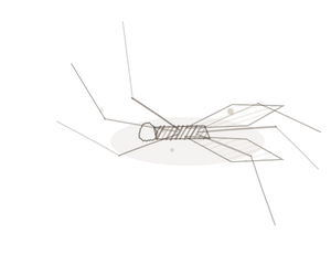
Keying out a specimen
Four questions, in this order
This is the procedure. It works on a slide you have never seen before, and it works best if you refuse to guess the answer before you have asked all four.
- Is the matrix solid? No → connective tissue proper, go to 2. Yes → supporting, go to 3.
- More space than fibre, or more fibre than space? Space → loose: empty circles mean adipose, a silver net means reticular, otherwise areolar. Fibre → dense: go to 4.
- Is it mineralised? Yes → bone: rings mean compact, struts mean spongy. No → cartilage: featureless matrix means hyaline, a dark mesh means elastic, thick bundles with cells in rows means fibrocartilage.
- Do the bundles agree about direction? All one way → dense regular. Every way at once → dense irregular. Wavy, dark and branching → elastic connective tissue.
One override: if what you have is a band of cartilage in stacked columns running down into new bone, you are not looking at a tissue at all. You are looking at the epiphyseal plate, and it is Pl. XII.
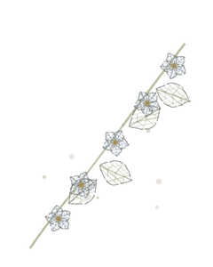
YOur missions
Field Notes for Future Missions
Before you go to the plates, try the key on a specimen nobody has named for you.
Mission 1 · how much is fibre
Mission 2
Field notes
Why twelve organisms
Connective tissue is proportion, and proportion is hard to teach from a picture because every micrograph is pink. The twelve organisms in this volume are each an argument about proportion made at a scale you can see without a microscope.
Spanish moss is mostly air. A honeypot ant is mostly cargo. A weaver nest has no plan and cannot be pulled apart; a hank of flax has nothing but a plan and frays if you cut across it. A coconut husk exists to be dropped. A reef is strong because it is mostly holes. Hold the organismA living individual made up of cells. in your headRounded proximal end that fits into the acetabulum of the hip bone. and the micrograph stops being pink and starts being an answer.
Begin · Pl. I
Laxus vagus var. tillandsius
the packing peanut · areolar connective tissue.
Hop to:
← Epithelial Tissues: A Guided Journey
All plates — The Tissue Field Guide: Epithelium
Frontispiece Ordo Epithelia (main page)
Pl. I · Simple squamous
Pl. II · Simple cuboidal
Pl. III · Simple columnar
Pl. IV · Stratified squamous
Pl. V · Stratified cuboidal
Pl. VI · Stratified columnar
Pl. VII · Transitional
Pl. VIII · Pseudostratified columnar
Epithelial Identification Game
All Modules
- Anatomical Language, Membranes & Homeostasis
- Just Enough Chemistry
- The Cell & Its Transport
- Making Cells & Proteins
- The Integumentary System
- The Skeletal System
- The Muscular System
- Nervous Tissue & the Senses
- The Spinal Cord
- The Brain & the Blood–Brain Barrier
- The Autonomic Nervous System
- The Special Senses
- The Endocrine System
- Blood
- The Heart
- Blood Vessels
- The Digestive System
- The Respiratory System
- The Urinary System
- Fluids, Electrolytes & Acid–Base Balance
- The Reproductive System
- The Immune System
List of terms
- hyaline cartilage
- elastic cartilage
- fibrocartilage
- compact bone
- spongy bone
- epiphyseal plate
- cells
- water
- molecules
- collagen
- cartilage
- chondrocyte
- osteocyte
- lacuna
- central canal
- dermis
- nuclei
- organism
- head