Time To Read
Date Last Modified
Frontispiece · ORD. NERVOSA
Ordo Nervosa
the wiring · nervous tissue
The plates
The six specimens
Six specimens, and nearly every one of them is told apart by pattern rather than by cell. Each plate gives you where the tissue lives, how to know it on sight, the look-alikes that catch people out, and a virtual slide to try the key on.

Pl. II
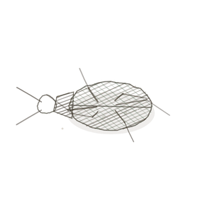
Periphericus globosus var. allianus
the garlic bulb · a ganglionA cluster of neuron cell bodies located in the peripheral nervous system (PNS).

Pl. III
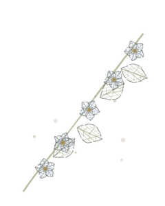
Centralis trilaminatus var. thujanus
the tree of life · cerebellumThe brain region responsible for balance, coordination, and fine motor control.

Pl. IV
Centralis corticalis var. lichenosus
the crust · cerebrumThe largest part of the brain, responsible for thought, memory, and voluntary movements.

Pl. V
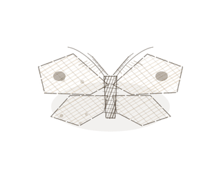
Centralis inversus var. interrogationis
the butterfly · the spinal cordThe central nervous system structure that relays signals between the brain and body.

Pl. VI
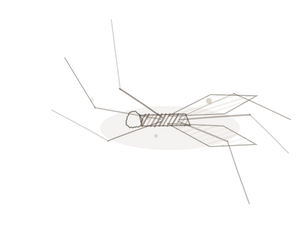
Centralis trimembranatus var. arachnoideus
the three veils · the meninges(singular: meninx) – Protective membranes surrounding the spinal cord and brain.

The ground plan
Two kinds of grey
Nervous tissue comes in exactly two textures, and almost everything else follows from knowing which one you are looking at. Where the cell bodies are, the tissue is crowded with nucleiClusters of neurons in the CNS responsible for processing information. and stains darker: grey matter. Where the myelinated axons are, it is pale, fibrous and nearly free of nuclei: white matterThe outer portion of the spinal cord made of myelinated nerve fibers that transmit signals.. Learn to see that difference and the rest of this volume is only asking where each one sits.
Fig. 1 · what makes grey grey

Fibre on one side, cell bodies on the other, and a surprisingly abrupt edge between them.
The five words
- Neuron — one nerve cell: a cell bodyThe central part of a neuron containing the nucleus and organelles., a tree of dendritesBranch-like extensions from a neuron that receive signals from other neurons., and a single long axon.
- Axon — that single long process. Most of what fills white matter is axons, not cellsThe basic structural and functional units of life..
- Myelin — the fatty wrapping around an axon. It is what makes white matter white.
- Grey matter — where the cell bodies are. Crowded nuclei, so it stains darker.
- White matter — where the myelinated axons are. Pale, fibrous, almost no nuclei.
Decide whether you are looking at cell bodies or at fibre before you say anything else out loud. Nearly every wrong answer in this volume begins by skipping that.

First question
Central or peripheral?
Everything here is either inside the brain and cord or outside them. The two have different rules, different cells doing the wrapping, and different answers to every later question — so ask this one first.
Fig. 2 · outside and in

The cord in its coverings, with a nerve leaving on each side. The boundary is right there.
- Peripheral — nerves and ganglia. Axons wrapped in sheaths, cell bodies kept in tidy capsules outside the cord.
- Central — brain, cerebellum, cord. No sheaths of that kind, and the cell bodies sit in sheets or horns rather than capsules.
- A capsule of big round cell bodies with a nerve coming out of it means peripheral, and you are nearly finished.
Peripheral splits fast: axons only means a nerve, cell bodies means a ganglion. Central needs one more question.
Second question
Where is the grey?
This is the question the whole volume turns on, and it has only two answers.
Fig. 3 · the inversion

A candy bar turned inside out — grey in the middle, pale rim around it. The brain does the opposite.
- Grey on the outside → brain. Cerebrum or cerebellum, and the layering tells you which.
- Grey on the inside → spinal cord. A butterfly of grey with white matter all around it.
Grey out, grey in. If you keep one sentence from this frontispiece, keep that one.

The naming key
How to read a name
Put the two answers together — where it lives first, then the pattern you can see — and you have said the name. The Latin on each plate is built the same way round.
First word · where it lives
- Periphericus — outside the brain and cord
- Centralis — inside them
Second word · the pattern
- fasciculatus — gathered into bundles (a nerve)
- globosus — round cell bodies in a capsule (a ganglion)
- trilaminatus — three layers (cerebellum)
- corticalis — a cortex, grey on the outside (cerebrum)
- inversus — grey on the inside instead (the cord)
- trimembranatus — three membranes (the meninges)
Worked both ways
- Centralis trilaminatus → central, three layers → cerebellum.
- Periphericus globosus → peripheral, round cell bodies → a ganglion.
- You saw grey in the middle of a pale rim → the cord → Centralis inversus.
Two that trip people up
The look-alikes
Neither of these breaks the key. They catch people out because the pair looks alike at low power, and the thing that separates them is small and specific.
Fig. 4 · the row that settles it
Cerebellum. One row of giant cells, exactly one cell deepAway from the surface of the body. — the cerebrum has nothing like it.
Cerebellum mistaken for cerebrum
Both are folded cortex with grey on the outside.
At low power both are a darker band following a folded surface over a pale core. Go up in power and look for a single row of enormous cells with flattened dendritic trees. If the row is there it is cerebellum, and the dense dark granular layer underneath will confirm it.
Find the Purkinje row, or establish that there is not one.
A nerve mistaken for a tendon
Both are pink, fibrous and full of long parallel things.
Cut across, a nerve is unmistakable: pale rings of myelin each with a dark axon at the centre, gathered into fasciclesBundles of nerve fibers within a nerve.
or
Bundles of nerve fibers within a muscle. with a sheath around each. Tendon cut across is solid pink with flattened nuclei squeezed between the bundles, and no rings at all.
Look for rings. Rings mean nerve.
Keying out a specimen
Four questions, in this order
This is the procedure. It works on a slide you have never seen before, which is the only kind that counts in a practical.
- Central or peripheral? A capsule of round cell bodies, or axons in sheaths → peripheral, go to 2. Otherwise → go to 3.
- Cell bodies or axons? Big round cell bodies each ringed by satellite nuclei → a ganglion. Rings of myelin and no cell bodies → a nerve.
- Where is the grey? Outside → brain, go to 4. Inside, as a butterfly → the spinal cord, and you are done.
- Is there a single row of giant cells? Yes → cerebellum. No, only a band that fades → cerebrum.
One override: if what you have is three membranes wrapped around nervous tissue rather than the tissue itself, you are on the meninges — and you name those by texture, outside in.
YOur missions
Field Notes for Future Missions
Before you go to the plates, try the key on a specimen nobody has named for you.
Mission 1 · grey out, grey in
Mission 2
Field notes
Why six organisms
Nervous histology is pattern recognition and very little else. There is no stain that names these for you and no single cell you can point at; there is only the arrangement — and arrangement is exactly the sort of thing a name slides straight off. So each specimen here is pinned to an organismA living individual made up of cells. whose own arrangement is the same idea in a form you cannot un-see.
A leek peels in order. A garlic bulb wraps every clove separately. A cedar branches the same way at every scale. A lichen is a crust with a cortex. A butterfly is known by its outline and not its colour. And a cellar spider’s web holds a space open rather than filling it. Those are the six, and you have all of them now.
Begin · Pl. I
Periphericus fasciculatus var. porraceus
the cable bundle · peripheral nerve.
Hop to:
← Epithelial Tissues: A Guided Journey
All plates — The Tissue Field Guide: Epithelium
Frontispiece Ordo Epithelia (main page)
Pl. I · Simple squamous
Pl. II · Simple cuboidal
Pl. III · Simple columnar
Pl. IV · Stratified squamous
Pl. V · Stratified cuboidal
Pl. VI · Stratified columnar
Pl. VII · Transitional
Pl. VIII · Pseudostratified columnar
Epithelial Identification Game
All Modules
- Anatomical Language, Membranes & Homeostasis
- Just Enough Chemistry
- The Cell & Its Transport
- Making Cells & Proteins
- The Integumentary System
- The Skeletal System
- The Muscular System
- Nervous Tissue & the Senses
- The Spinal Cord
- The Brain & the Blood–Brain Barrier
- The Autonomic Nervous System
- The Special Senses
- The Endocrine System
- Blood
- The Heart
- Blood Vessels
- The Digestive System
- The Respiratory System
- The Urinary System
- Fluids, Electrolytes & Acid–Base Balance
- The Reproductive System
- The Immune System
List of terms
- ganglion
- cerebellum
- cerebrum
- spinal cord
- meninges
- nuclei
- white matter
- cell body
- dendrites
- cells
- deep
- fascicles
- organism